


Research Article
Open Access
Preparation of Biocompatible Palladium-Fe3O4
Nanoparticles/Multiwalled Carbon Nanotubes Composite
and its Electrocatalytic Activity towards Determination of
Cholesterol on Screen Printed Electrode
Revanasiddappa Manjunatha1, Gurukar S. Suresh1, 2*, Jose S. Melo3, 4*,
Jakkid Sanetuntikul5 and Sangaraju Shanmugam5
1Chemistry Research Centre, S. S. M. R. V. Degree College, Jayanagar, Bangalore - 560041, India
2Department of Chemistry and Research Centre, N.M.K.R.V. College for Women, Jayangar, Bangalore -560011, India
3Nuclear Agriculture and Biotechnology Division, Bhabha Atomic Research Centre, Trombay, Mumbai - 400085, India
4Homi Bhabha National Institute, Anushakti Nagar, Mumbai- 400094, India
5Department of Energy Systems and Engineering, Daegu Gyeongbuk Institute of Science and Technology, Daegu 711-873, Republic of Korea
2Department of Chemistry and Research Centre, N.M.K.R.V. College for Women, Jayangar, Bangalore -560011, India
3Nuclear Agriculture and Biotechnology Division, Bhabha Atomic Research Centre, Trombay, Mumbai - 400085, India
4Homi Bhabha National Institute, Anushakti Nagar, Mumbai- 400094, India
5Department of Energy Systems and Engineering, Daegu Gyeongbuk Institute of Science and Technology, Daegu 711-873, Republic of Korea
*Corresponding author: Jose Savio Melo, Nuclear Agriculture and Biotechnology Division, Bhabha Atomic Research Centre, Trombay and HomiBhabha National Institute, Anushakti Nagar, Mumbai- 400 085, India, Telephone: 91-22-25592760; Fax no: 91-22-25505151.;
E-mail:
@
Gurukar S. Suresh, Department of Chemistry and Research Centre, N.M.K.R.V. College for Women, Jayangar, Bangalore -560011, India, Telephone: 91 –80 – 26654920;Fax no: 91 – 80 – 22453665 ; E-mail:@
Gurukar S. Suresh, Department of Chemistry and Research Centre, N.M.K.R.V. College for Women, Jayangar, Bangalore -560011, India, Telephone: 91 –80 – 26654920;Fax no: 91 – 80 – 22453665 ; E-mail:
Received: 03 October, 2016; Accepted: 04 January, 2017; Published: 10 February, 2017
Citation: Jose S. Melo (2017) Preparation of Biocompatible Palladium-Fe3O4 Nanoparticles/Multiwalled Carbon Nanotubes Composite and its Electrocatalytic Activity towards Determination of Cholesterol on Screen Printed Electrode. J of Biosens Biomark Diagn
2(1):1-10. DOI: 10.15226/2575-6303/2/1/00109
Abstract
A simple and facile microwave method was adopted to prepare
Fe3O4 and Pd- Fe3O4 nanoparticles, which possess the mean particle
diameter of 10 nm and 90 nm, respectively. Formation of Fe3O4 and Pd-
Fe3O4 nanoparticles were confirmed from Powder X-ray diffraction,
Transmission electron microscopy, Energy-dispersive X-ray
spectroscopy, and FT-Infra red spectroscopy techniques. Negatively
charged mutliwalled carbon nanotubes (COO--MWCNTs) were
wrapped with positively charged poly (Diallyldimetheylammonium
Chloride) (PDDA) followed by coating with Pd-Fe3O4 nanoparticle
to get (Pd-Fe3O4/PDDA/COO- -MWCNTs) composite. This composite
was used for the determination of cholesterol by using Cholesterol
Oxidase (ChOx) enzyme on Screen Printed Electrode (SPE). (Pd-
Fe3O4/PDDA/COO- -MWCNTs) composite provides biocompatible
microenvironment for the ChOx to exhibit Direct Electron Transfer
(DET) on electrode surface. A well defined redox peak at -0.365 and
-0.443 V was observed, corresponding to the DET of the FAD/FADH2 of
ChOx. Enzyme modified SPE was characterized by cyclic voltammetry
and electrochemical impedance spectroscopy by means of Fe(CN)6 3-/4as an electrochemical probe. The linear range of the enzyme modified
SPE was found to be 10-80 μm (R=0.9972) with detection limit of 1 μm
of cholesterol. The sensitivity of the enzyme modified SPE was found
to be 10.45 μA μM-1 cm-2 for the determination of cholesterol, common
interferents such as ascorbic acid, uric acid and glucose did not cause
any interference because of low operating potential.
Keywords: Pd-Fe3O4; Nanoparticles; Screen Printed Electrode; Cholesterol Oxidase
Keywords: Pd-Fe3O4; Nanoparticles; Screen Printed Electrode; Cholesterol Oxidase
Introduction
Preparation of Biocompatible Palladium-Fe3O4
Nanoparticles/Multiwalled Carbon Nanotubes
Composite and its Electrocatalytic Activity towards
Determination of Cholesterol on Screen Printed
Electrode
The magnetic nanoparticles in general, iron oxide
(Fe3O4) have been attracted an increasing interest in the
development of nanostructured materials and nanotechnology in
biotechnology and medicine [1]. The main advantage of magnetic
nanoparticles is that, they can easily and rapidly separate
from their matrix by an external magnetic field. Some of the
common features associated with the Fe3O4 nanoparticles are
good biocompatibility, strong superparamagnetic property, high
surface area, low toxicity and ease of preparation [2,3]. Thus,
Fe3O4 nanoparticles have been used in a wide range of potential
applications such as, electrochemical sensors/biosensors,
catalysis, immunoassays, data storage [4-7]. Various methods
have been used for the synthesis of magnetic nanoparticles
which includes hydrothermal synthesis, co-precipitation, sol-gel
method, microwave irradiation method [8-11]. The later method
has significant advantage with respect to higher reaction rates
and product yields in a shorter period of time.
Carbon nanotubes are one of the new kinds of carbon material, discovered in the last decades of the 20th century [12]. Researchers have explored various potential characteristics of CNTs, which could be applicable to various fields [13]. It was found that CNTs have excellent electronic conductivity, mechanical strength, chemical stability and unique structural properties [14]. However, the main drawbacks exist in the processibility of CNTs in solution, in which they precipitate into ropes or bundles due to strong Van der Waals interactions [15]. To overcome this problem, surface modifications have been employed, such as chemical functionalization using strong acids, polymer wrapping of CNTs [16,17]. In later method i.e. chemical functionalization of CNTs using strong acid results in partial oxidation of the carbon atoms to produce oxygen containing groups such as carboxylic groups, especially in the open ends of CNTs [18]. These groups are negatively charged in the aqueous solution and can interact with positively charged poly electrolytes [19].
Cholesterol is one of the most important analyte in clinical analysis, because its assay is important for diagnosis and prevention of a numerous clinical disorders such as, hypertension, cerebral thrombosis, arteriosclerosis and coronary heart disease [20]. Recent studies explored that cholesterol plays a vital role in the brain synapses and also in the immune system including protection against cancer. In earlier days, cholesterol was determined by using non-enzymatic spectrophotometric techniques by using colored substances [21]. However, this technique suffers from low specificity, instability of reagents and high cost. These can be effectively addressed by using enzymatic cholesterol biosensor. Some of the advantages of enzymatic cholesterol biosensors are specificity, simplicity, rapidness and cost effectiveness [22]. The most commonly used enzyme for the construction of cholesterol biosensor is cholesterol oxidase. ChOx is a Flavin-Adenine-Dinucleotide (FAD) containing flavoenzyme. In the presence of oxygen, ChOx catalyzes two reactions; oxidation of cholesterol to cholest-5-en-3-one and subsequently the isomerization to cholest-4-en-3-one.
Direct electrochemistry of redox enzyme systems has gained increasing interest both for the study of the electron transport proteins as well as development of third generation reagent less electrochemical biosensors [23,24]. However, direct electron transfer between redox enzyme and electrode is generally difficult to observe due to several factors. Such as, enzyme active sites are deeply embedded in protein matrix, resulting in a long distance between active sites and underlying electrode. In addition, conformational changes or denaturation of redox enzyme often occur while immobilization of enzyme onto the electrode surface. Thus, to obtain DET, many techniques have been developed such as layer-by-layer technique, covalent binding using cross linkers, physical adsorption [25-27]. Physical adsorption method involves van der Waals forces, ionic binding or hydrophobic forces. The main advantage of this method is that it is simple and can be used under mild conditions. It requires only a minimum activation steps resulting in little or no conformational changes of the enzyme or destruction of its active centre [28].
In the present work, a simple and facile microwave method was adopted to prepare Fe3O4 and Pd- Fe3O4 nanoparticles by following the procedure given in the research article with slight modifications [29]. Formation of both Fe3O4 and Pd-Fe3O4 nanoparticles were confirmed by pXRD, TEM, EDX and FTIR analysis. Negatively charged Pd- Fe3O4 nanoparticles were mixed with positively charged PDDA wrapped MWCNTs to get negatively charged novel composite. This negatively charged novel composite was drop casted on the Screen Printed Electrode (SPE). Positively charged ChOx (pH 4. 0) was immobilized on the composite by physical adsorption method. Enzyme modified screen printed electrode showed electrocatalytic activity towards detection of cholesterol. Cyclic Voltammery (CV) and Electrochemical Impedance Spectroscopy (EIS) techniques were used to characterize the enzyme modified screen printed electrode. Above mentioned novel composite provides biocompatible microenvironment for the ChOx to exhibit DET on SPE. To the best of our knowledge, for the first time we have used this novel composite for detection of cholesterol.
Carbon nanotubes are one of the new kinds of carbon material, discovered in the last decades of the 20th century [12]. Researchers have explored various potential characteristics of CNTs, which could be applicable to various fields [13]. It was found that CNTs have excellent electronic conductivity, mechanical strength, chemical stability and unique structural properties [14]. However, the main drawbacks exist in the processibility of CNTs in solution, in which they precipitate into ropes or bundles due to strong Van der Waals interactions [15]. To overcome this problem, surface modifications have been employed, such as chemical functionalization using strong acids, polymer wrapping of CNTs [16,17]. In later method i.e. chemical functionalization of CNTs using strong acid results in partial oxidation of the carbon atoms to produce oxygen containing groups such as carboxylic groups, especially in the open ends of CNTs [18]. These groups are negatively charged in the aqueous solution and can interact with positively charged poly electrolytes [19].
Cholesterol is one of the most important analyte in clinical analysis, because its assay is important for diagnosis and prevention of a numerous clinical disorders such as, hypertension, cerebral thrombosis, arteriosclerosis and coronary heart disease [20]. Recent studies explored that cholesterol plays a vital role in the brain synapses and also in the immune system including protection against cancer. In earlier days, cholesterol was determined by using non-enzymatic spectrophotometric techniques by using colored substances [21]. However, this technique suffers from low specificity, instability of reagents and high cost. These can be effectively addressed by using enzymatic cholesterol biosensor. Some of the advantages of enzymatic cholesterol biosensors are specificity, simplicity, rapidness and cost effectiveness [22]. The most commonly used enzyme for the construction of cholesterol biosensor is cholesterol oxidase. ChOx is a Flavin-Adenine-Dinucleotide (FAD) containing flavoenzyme. In the presence of oxygen, ChOx catalyzes two reactions; oxidation of cholesterol to cholest-5-en-3-one and subsequently the isomerization to cholest-4-en-3-one.
Direct electrochemistry of redox enzyme systems has gained increasing interest both for the study of the electron transport proteins as well as development of third generation reagent less electrochemical biosensors [23,24]. However, direct electron transfer between redox enzyme and electrode is generally difficult to observe due to several factors. Such as, enzyme active sites are deeply embedded in protein matrix, resulting in a long distance between active sites and underlying electrode. In addition, conformational changes or denaturation of redox enzyme often occur while immobilization of enzyme onto the electrode surface. Thus, to obtain DET, many techniques have been developed such as layer-by-layer technique, covalent binding using cross linkers, physical adsorption [25-27]. Physical adsorption method involves van der Waals forces, ionic binding or hydrophobic forces. The main advantage of this method is that it is simple and can be used under mild conditions. It requires only a minimum activation steps resulting in little or no conformational changes of the enzyme or destruction of its active centre [28].
In the present work, a simple and facile microwave method was adopted to prepare Fe3O4 and Pd- Fe3O4 nanoparticles by following the procedure given in the research article with slight modifications [29]. Formation of both Fe3O4 and Pd-Fe3O4 nanoparticles were confirmed by pXRD, TEM, EDX and FTIR analysis. Negatively charged Pd- Fe3O4 nanoparticles were mixed with positively charged PDDA wrapped MWCNTs to get negatively charged novel composite. This negatively charged novel composite was drop casted on the Screen Printed Electrode (SPE). Positively charged ChOx (pH 4. 0) was immobilized on the composite by physical adsorption method. Enzyme modified screen printed electrode showed electrocatalytic activity towards detection of cholesterol. Cyclic Voltammery (CV) and Electrochemical Impedance Spectroscopy (EIS) techniques were used to characterize the enzyme modified screen printed electrode. Above mentioned novel composite provides biocompatible microenvironment for the ChOx to exhibit DET on SPE. To the best of our knowledge, for the first time we have used this novel composite for detection of cholesterol.
Methods
Reagents
TritonX-100,Cholesterol, Pd(NH3)4Cl2•H2O, FeSO4•7H2O,
MWCNTs and PDDA (Mw: 200,000-350,000), were purchased
from Sigma Aldrich. Cholesterol oxidase was procured from SRL,
India. Screen printed electrodes of diameter 3 mm (0.071 cm2)
were purchased from CH instruments (product no. TE 100).
Phosphate Buffer Saline of pH 7. 0 (PBS) was prepared from stock
solutions of 0.1 M KH2PO4, 0.1 M K2HPO4 and 0.1M KCl. All other
chemicals used were of analytical reagent grade unless otherwise
mentioned and used without further purification. All solutions
were prepared with milli-Q water.
Enzyme solution preparation
100 U/ml ChOx solution was prepared in 0.1 M acetate
buffer solution of pH 4. A stock solution of 10 mM cholesterol was
prepared by dissolving 0.0967 g of cholesterol in a mixture of 1
mL Triton X-100 and 0.5 mL isopropanol at 65°C and diluting the
resulting solution to 25 mL in a standard flask using hot PBS of
pH 7.0. The solution was stored at 4°C in the dark and was stable
for two weeks (until a slight turbidity was observed).
Synthesis of Fe3O4 and Pd- Fe3O4 nanoparticles
As stated earlier, Fe3O4 and Pd- Fe3O4 nanoparticles were
prepared according to the procedure described elsewhere [29]. In
brief, 2.0 mm FeSO4.7H2O was dissolved in 100 ml distilled water
with continuous stirring. The pH of the solution was adjusted to
11 by conc. ammonia solution resulting in the formation of black
precipitation. This suspension was transferred into microwave
oven, in which microwave radiation of high energy was applied
for one minute. The product Fe3O4 nanoparticles were separated
by centrifugation method. Finally Fe3O4 nanoparticles washed
thoroughly with water followed by ethanol and dried at 50°C in
vacuum oven.
Similarly, Pd- Fe3O4 nanoparticles were prepared by coprecipitation method. 0.5 mm Pd (NH3)4Cl2•H2O, and 2.0 mmol FeSO4•7H2O were dissolved in 100 ml distilled water with constant stirring. The pH of the solution was adjusted to about 11 by conc. ammonia solution. Then the reaction mixer was taken into microwave oven for microwave irradiation. The product was isolated by using above descried procedure.
Similarly, Pd- Fe3O4 nanoparticles were prepared by coprecipitation method. 0.5 mm Pd (NH3)4Cl2•H2O, and 2.0 mmol FeSO4•7H2O were dissolved in 100 ml distilled water with constant stirring. The pH of the solution was adjusted to about 11 by conc. ammonia solution. Then the reaction mixer was taken into microwave oven for microwave irradiation. The product was isolated by using above descried procedure.
Electrochemical measurements
Cyclic voltammetry, electrochemical impedance
spectroscopy, differential pulse voltammetry experiments were
carried out with Versa stat 3 (Princeton Applied Research, USA).
The microwave oven used in the present study was a domestic
microwave oven (LG, intellowave, MS-2342 AE). Powder X-Ray
Diffraction (pXRD) patterns of the samples were recorded using
a Philips X’pert Pro diffractometer with CuKα (λ = 1.5418 Ǻ).
FT-IR experiments were carried out with Bruker Alpha-T FTIR
spectrometer (ATR mode, diamond crystal, resolution 4 cm-1,
400-4000 cm-1). The morphology of the samples were analyzed
by the Field emission scanning electron microscopy (Hitachi,
S4800 FE-SEM) and the Field emission transmission electron
microscopy (Hitachi, HF 3600 FE-TEM). TEM experiments were
performed at an acceleration voltage of 300 kV. For the elemental
mapping study, an Energy Dispersive X-Ray Spectroscopy (EDXS)
connected to a TEM was used in scanning mode. TEM samples
were prepared by dropping ultrasonically dispersed isopropyl
alcohol solution of nanoparticles on a copper grid coated with
amorphous carbon film. All experiments were done in an
electrochemical cell consisting of SPE with an unmodified or
modified carbon working electrode, a carbon counter electrode
and Ag/AgCl reference electrode.
Preparation of enzyme modified screen printed
electrode
As we already discussed in the introduction, CNTs are insoluble
in most of the solvents because they precipitate into ropes are
bundles due to strong Vander Waals interactions. To overcome this
difficulty, we introduced carboxylic groups on MWCNTs surface
by refluxing with conc. nitric acid for 5 h, followed by filtration
and washed with pure water until the filtrate become neutral.
Finally the product was dried in vacuum at 50°C [30]. PDDA is a
water soluble, quaternary ammonium cationic polyelectrolyte. It
is positively charged colloid when dissolved in aqueous solutions
[31,32]. The positively charged PDDA polymer can be easily
wrapped/coated on negatively charged MWCNTs [33]. 1 mg/
ml carboxylated MWCNTs dispersed in 0.2% PDDA solution,
ultrasonicalted for 20 min. followed by stirred at 50°C for 12 h.
To this composite 0.5 mg/ml Pd- Fe3O4 nanoparticles were added
and stirred for 12 h at room temperature. In this stage negatively
charged Pd- Fe3O4 nanoparticles coated on positively charged
MWCNTs wrapped with PDDA as shown in figure 1.

Figure 1:Schematic representation for the fabrication of enzyme electrode
based on the (PdFe3O4/PDDA/COO- -MWCNTs) composite.
2.5 μl of Pd- Fe3O4/PDDA/COO- -MWCNTs composite was drop
casted on screen printed electrode, dried at ambient temperature.
5 μl of positively charged ChOx (pH 4.0) drop casted on composite,
dried at 4°C. Hereafter, the enzyme modified electrode was
denoted as ChOx-(Pd- Fe3O4/PDDA/COO- -MWCNTs)/SPE.
Results and discussion
Characterization of Fe3O4 and Pd- Fe3O4 nano particles
using pXRD, TEM, EDX and FT-IR techniques
Figure 2a shows XRD patterns of the synthesized Fe3O4
nanoparticles. Diffraction peaks of Fe3O4 nanoparticles were
obtained at 30.18°, 35.54°, 43.29°, 53.69°, 57.29° and 62.96°
corresponding to the index planes (220), (311), (400), (422),
(511) and (440) respectively. This is quite identical to pure Fe3O4
nanoparticles and well matched with that of JCPDS no. 82-1533.
This revealed that the Fe3O4 nanoparticles have a cubic spinel
structure [34,35]. Also, no characteristic peaks of impurities
were observed. From the XRD data, the mean particle diameter
of Fe3O4 nanoparticles was calculated from index planes (220),
(311) and (400) by using Debye-Schererrer’s equation, which is
given below.)

Figure 2:A. XRD patterns of Fe3O4 (a) and Pd- Fe3O4 (b) nanoparticles.
B. FT-IR spectrum of Fe3O4 nanoparticles.
Where, λ- wavelength of X-ray, β- full width at half
maximum,θ- Bragg’s diffraction angle. The mean particle diameter
of Fe3O4 nanoparticles was found to be 10 nm. These results
depicts that the Fe3O4 nanoparticles can be rapidly synthesized
with 5-10 minutes. Usually most of the Fe3O4 nanoparticles
synthetic methods need more than an hour [36].
Formation of Fe3O4 nanoparticles by microwave irradiation can be explained as follows
Formation of Fe3O4 nanoparticles by microwave irradiation can be explained as follows
When ammonia is added to the FeSO4 solution Fe(OH)2 is
formed according to the equation 2, which is oxidized to Fe3O4
nanoparticles und the influence of microwave radiation as follows
Similarly figure 2b shows XRD patterns of the Fe3O4
nanoparticles decorated on Pd. The addition diffraction peaks
at 40.07°, 46.54° and 68.09° corresponding to the (111), (2000)
and (220) lattice planes were attributed to formation of Pd
nanoparticles [37,38]. The mean particle diameter of Pd-Fe3O4
nanoparticles was found to 90 nm according to equation 1. The
possible mechanism for the formation of Pd-Fe3O4 shown as
below,
FT-IR data of Fe3O4 nanoparticles is shown in figure 2B.
It is noteworthy that in figure 2B, peak at 545 cm-1 is attributed
to the Fe-O bond vibration of Fe3O4 [39]. The broad peak at
3346 cm-1 is due to stretching vibrations of –OH bond, which
is absorbed by Fe3O4 nano particles. Also, the peak at ∼1610
cm-1 may be assigned to the deformation vibrations of water
molecules trapped onto the magnetic nanoparticles [40]. These
results confirm the formation of Fe3O4 nanoparticles. There was
no major change in the FT-IR spectrum of Pd-Fe3O4 (results not
shown).


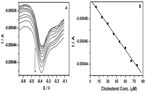
Figure 3:(a) TEM (b) HRTEM images of Fe3O4 nanoparticles. B. (a) TEM
(b) HRTEM Images of Pd-Fe3O4 nanoparticles. C. (a)
Bright field TEM image Pd-Fe3O4 nanoparticles, the corresponding EDX
maps depict the distribution of constituting individual elements within
the structure as shown in (b-d). The images correspond to the (b) Fe,
(c) Pd, and (d) O.
Figure 3A, 3B shows TEM images of Fe3O4 and Pd-
Fe3O4 nanoparticles respectively. Both Fe3O4 and Pd- Fe3O4
nanoparticles appeared to be almost spherical in shape. TEM
image of figure 3B (a) clearly depicts the presence of slightly
bigger Pd nanoparticles (dark contrast) surrounded by Fe3O4
nanoparticles, which is evidenced in the difference in contrast.
The average diameter of Fe3O4 nanoparticles was found to be 12.8
nm, whereas, the average particle size of Pd- Fe3O4 nanoparticles
was found to be 92.4 nm. These results are in well agreement with
XRD results shown in figure 3A. The high resolution TEM (HRTEM)
images of Fe3O4 and Pd-Fe3O4 nanoparticles are shown in figure
3A (b) & 3B (b), respectively. The distance between two lattice
planes of the Fe3O4 crystallite was 0.252 nm, which corresponds
to the (311) plane of spinal Fe3O4. In the same way, HRTEM image
of Pd- Fe3O4 nanoparticle showed well resolved lattice fringes
with a distance of 0.225 nm, corresponding to the (111) plane
of cubic Pd. Furthermore, the presence of Pd nanoparticles in
Fe3O4 was also confirmed using scanning transmission electron
microscope (STEM) coupled with elemental mapping analysis
(STEM-EDS). Figure 3C (a) shows a bright field TEM image of
Pd-Fe3O4 nanoparticles. The corresponding elemental maps are
given in figure 3C (b-d). The EDS mapping results suggest that
metallic Pd core is surrounded by several Fe3O4 nanoparticles.
Figure 4:SEM images of unmodified SPE (a), (PDDA/COO- -MWCNTs)/
SPE (b), (Pd-Fe3O4/PDDA/COO- -MWCNTs)/SPE(c)andChOx-(Pd-Fe3O4/
PDDA/COO- -MWCNTs)/SPE (d).
Figure 4 displays the SEM images of unmodified SPE
(a), (PDDA/COO- -MWCNTs)/SPE (b), (Pd-Fe3O4/PDDA/COO- -MWCNTs)/SPE (c) and ChOx-(Pd- Fe3O4/PDDA/COO- -MWCNTs)/
SPE (d). The morphology of the unmodified SPE exhibits rough
surface having different size grains of several microns. PDDA
coated MWCNTs composite is uniformly deposited on SPE, which
can be seen in image (b). Nano sized Pd-Fe3O4 particles appear
as small white grains which are incorporated into (PDDA/COO- -MWCNTs) composite clearly visible in image (c). Image (d)
shows uniform immobilization of ChOx enzyme on (Pd-Fe3O4/
PDDA/COO- -MWCNTs) composite. Inset shows the different size
granules of ChOx enzyme.
Characterization of ChOx-(Pd- Fe3O4/PDDA/COO- -MWCNTs)/SPE using CV and EIS
Fe(CN)6 3-/4- redox couple is widely used as an
electrochemical probe to characterize the property of
unmodified/modified electrodes. Figure 5
Figure 5:Cyclic voltammograms of unmodified SPE (a), Pd- Fe3O4/
PDDA/COO- -MWCNTs)/SPE (b) and ChOx-(Pd- Fe3O4/PDDA/COO- -MWCNTs)/
SPE (c) in 0.1 M PBS containing 5 mM Fe(CN)64−/3− (pH 7.0); scan
rate: 50 mVs-1.
Illustrates, the cyclic volt ammogramms of SPE, (Pd-
Fe3O4/PDDA/COO- -MWCNTs)/SPE and ChOx-(Pd-Fe3O4/PDDA/
COO- -MWCNTs)/SPE in 5 mM Fe(CN)63-/4- containing PBS (pH
7.0) at scan rate of 50 mVs-1. Irreversible voltammogram was
observed at SPE (curve a, dotted line) with minimal cathodic
peak current (Ipc) and anodic peak current (Ipa). The cathodic
peak potential (Epc) and anodic peak potential (Epa) were
found at 521 mV and -256 mV respectively with peak to peak
separation (ΔEp) 777 mV. However, SPE modified with (Pd-
Fe3O4/PDDA/COO- -MWCNTs) composite, a well redox peak of
Fe(CN)63-/4- with the ΔEp 48 mV (curve b) was obtained. These
results depict that the over potential decreased by 729 mV and
around 2 fold of increase in current was observed for 5 mM
Fe(CN)63-/4- at (Pd- Fe3O4/PDDA/COO- -MWCNTs)/SPE than that
of SPE. This shows that Electrocatalytic activity of (Pd- Fe3O4/
PDDA/COO- -MWCNTs) composite. After immobilization of ChOx
enzyme on (Pd- Fe3O4/PDDA/COO- -MWCNTs)/SPE, decreased in
peak current was observed (curve c). This could be attributed to
macromolecular non conducting enzymatic structure, impede
electrochemical redox reaction of Fe(CN)63-/4 -at electrode surface.
This demonstrates that ChOx enzyme successfully immobilized
on (Pd- Fe3O4/PDDA/COO- -MWCNTs) composite by means of
electrostatic attraction.
Elctrochemical Impedance Spectroscopy (EIS) is a powerful and sensitive characterization tool for studying the charge transfer process at electrode/electrolyte interface [41]. Hence, Characterization of SPE, (Pd- Fe3O4/PDDA/COO- -MWCNTs)/SPE and ChOx-(Pd- Fe3O4/PDDA/COO- -MWCNTs)/SPE was further investigated using EIS. EIS was carried out in the presence of 5 mM Fe (CN)64−/3−as a electrochemical redox probe, in the frequency range of 100 kHz to 0.1 Hz with amplitude of 5 mV as shown in figure 6A The equivalent circuit shown in the inset of figure 6B was used to fit experimental data. The simulated curve of experimental data and best fitting equivalent circuit are shown in the figure 6C. The obtained impedance data are shown in Table 1.
The circuit includes the solution resistance (Rs), charge transfer resistance (Rct), double layer capacitance (Qdl), Warburg impedance (Zw), Faradaic resistance, (Rf) and Faradaic capacitance (Cf). At SPE, big semicircle having a Rct of 5714 Ω was observed for Fe(CN)64−/3− (curve a), which suggests that unmodified SPE exhibits sluggish and unfavorable Fe(CN)64−/3− electron transfer. However, SPE modified with (Pd- Fe3O4/
Elctrochemical Impedance Spectroscopy (EIS) is a powerful and sensitive characterization tool for studying the charge transfer process at electrode/electrolyte interface [41]. Hence, Characterization of SPE, (Pd- Fe3O4/PDDA/COO- -MWCNTs)/SPE and ChOx-(Pd- Fe3O4/PDDA/COO- -MWCNTs)/SPE was further investigated using EIS. EIS was carried out in the presence of 5 mM Fe (CN)64−/3−as a electrochemical redox probe, in the frequency range of 100 kHz to 0.1 Hz with amplitude of 5 mV as shown in figure 6A The equivalent circuit shown in the inset of figure 6B was used to fit experimental data. The simulated curve of experimental data and best fitting equivalent circuit are shown in the figure 6C. The obtained impedance data are shown in Table 1.
The circuit includes the solution resistance (Rs), charge transfer resistance (Rct), double layer capacitance (Qdl), Warburg impedance (Zw), Faradaic resistance, (Rf) and Faradaic capacitance (Cf). At SPE, big semicircle having a Rct of 5714 Ω was observed for Fe(CN)64−/3− (curve a), which suggests that unmodified SPE exhibits sluggish and unfavorable Fe(CN)64−/3− electron transfer. However, SPE modified with (Pd- Fe3O4/
Table 1: EIS data of unmodified SPE, (Pd- Fe3O4/PDDA/COO- -MWCNTs)/SPE
and ChOx-(Pd- Fe3O4/PDDA/COO- -MWCNTs)/SPE in 5 mM Fe(CN)6 3-/4
Electrode |
Rs /Ω |
n |
Q/µF |
Rct /Ω |
W /Ω |
SPE |
189 |
0.95 |
0. 49 |
5714 |
0.0009 |
Pd- Fe3O4/PDDA/ |
198 |
0.97 |
0. 23 |
0.19 |
0.006 |
ChOx-(Pd- Fe3O4/PDDA/ COO--MWCNTs)/SPE |
196 |
0.78 |
14.5 |
557 |
0.0016 |
Figure 6:Nyquist impedance plots of unmodified SPE (a), Pd- Fe3O4/
PDDA/COO- -MWCNTs)/SPE (b) and ChOx-(Pd- Fe3O4/PDDA/COO- -MWCNTs)/
SPE (c) in 0.1 MPBS containing 5 mM Fe(CN)64−/3− (pH 7.0). The frequency range is from 100 kHz to 0.1 Hz and amplitude 5 mV. B. The
equivalent circuit used to fit experimental data. (C) Nyquist impedance
plots of experimental data of ChOx-(Pd-Fe3O4/PDDA/COO- -MWCNTs)/
SPE and best fitting by using equivalent.
PDDA/COO- -MWC) composite, big semicircle was
replaced with straight line, which have a Rct of 0.19 Ω as shown
in figure 5A inset (curve b). This shows (Pd- Fe3O4/PDDA/COO- -MWCNTs) composite facilitates fast and favorable electron
transfer towards electrode surface. Small semicircle was observed
(curve c) with a Rct of 557 Ω, when ChOx enzyme immobilized
on (Pd- Fe3O4/PDDA/COO- -MWCNTs)/SPE, which illustrates
that non conducting macromolecular ChOx was successfully
immobilized modified SPE and it oppose the electron transfer
towards electrode surface.
Direct electrochemistry of ChOx on (Pd- Fe3O4/PDDA/
COO- -MWCNTs)/SPE
Figure 7 shows, cyclic voltammogram of ChOx on
unmodified SPE (curve a) and (Pd- Fe3O4/PDDA/COO- -MWCNTs)/
SPE (curse b). When ChOx immobilized on unmodified SPE,
which does not facilitate DET. This illustrates that unmodified
SPE does not provide micro environment for the immobilization
of ChOx. However, ChOx immobilized on (Pd-Fe3O4/PDDA/COO- -MWCNTs)/SPE exhibits a pair of well defined redox peaks at
-0.365 and -0.443, these peaks are assigned for FAD/FADH2,
which could be ascribed to electron transfer between ChOx
and under laying electrode [19,22,27]. The potential difference
between the two peaks ΔEp was 78
Figure 7: Cyclic voltammograms of ChOx-unmodified SPE (a) and ChOx-
(Pd-Fe3O4/PDDA/COO- -MWCNTs)/SPE in 0.1M PBS (pH 7.0) scan rate:
50mVs-1.
mV at a scan rate of 50 mVs-1, which suggests that ChOx has
undergone a quasi-reversible redox reaction (Pd-Fe3O4/PDDA/
COO- -MWCNTs)/SPE. The surface area (τ) of ChOx-(Pd-Fe3O4/
PDDA/COO- -MWCNTs)/SPE was calculated using the following
equation.
Where, Q is the charge, n is the number of electrons
transferred, F is the Faraday constant. Therefore (τ) was found to
be 5.38 × 10⁻⁹ mol cm-2.
Effect of scan rate and pH at ChOx-(Pd-Fe3O4/PDDA/
COO- -MWCNTs)/SPE
To determine the kinetics of electrode reactions, the effect
of scan rate on the voltammetric response of ChOx-(Pd-Fe3O4/
PDDA/COO- -MWCNTs)/SPE in a 0.1 M PBS of pH 7 was studied
in the range of 5-75 mVs-1 as shown in figure 8A. The linear
regression equations are as given below
Ipa = -6.6628E-7 + 5.4642E-7 ν (Vs-1); R = 0.9981 (8)
Ipc = -1.4345E-5 - 6.3799E- 7 ν (Vs-1); R = 0.9932 (9)
Ipa = -6.6628E-7 + 5.4642E-7 ν (Vs-1); R = 0.9981 (8)
Ipc = -1.4345E-5 - 6.3799E- 7 ν (Vs-1); R = 0.9932 (9)
The redox peak current of ChOx increased linearly with
increasing scan rate (Figure 8B) and the peak to peak separation
also increased, indicating that surface controlled quasi-reversible
process is involved.
In addition, the anodic peak potential shifted to a more potential value with increasing scan rate, where as the cathodic peak potential shifted in a negative direction. The pH of the electrolyte solution has a significant influence on the redox reaction of FAD/FADH2 of ChOx with respect to peak current and peak potential. Figure 9A Shows cyclic voltammograms of effect of pH of electrolyte in the range 4 to 8 studied on the response of ChOx-(Pd-Fe3O4/PDDA/COO- -MWCNTs)/SPE. The electrochemical response of enzyme immobilized on the electrode surface is due to redox reaction of its active site, i.e. FAD/FADH2. Where, FAD is known to undergo redox reaction involving two electrons with two protons to form FADH2.The redox peak current of ChOx increased linearly with increasing scan rate (Figure 8B) and the peak to peak separation also increased, indicating that surface controlled quasi-reversible process is involved.
In addition, the anodic peak potential shifted to a more potential value with increasing scan rate, where as the cathodic peak potential shifted in a negative direction. The pH of the electrolyte solution has a significant influence on the redox reaction of FAD/FADH2 of ChOx with respect to peak current and peak potential. Figure 9A Shows cyclic voltammograms of effect of pH of electrolyte in the range 4 to 8 studied on the response of ChOx-(Pd-Fe3O4/PDDA/COO- -MWCNTs)/SPE. The electrochemical response of enzyme immobilized on the electrode surface is due to redox reaction of its active site, i.e. FAD/FADH2. Where, FAD is known to undergo redox reaction involving two electrons with two protons to form FADH2.The redox peak current of ChOx increased linearly with increasing scan rate (Figure 8B) and the peak to peak separation also increased, indicating that surface controlled quasi-reversible process is involved.
Figure 8: Cyclic voltammograms of ChOx-(Pd- Fe3O4/PDDA/COO- -MWCNTs)/
SPE in PBS (pH 7.0) at different scan rates: (5-75 mV-1). B. The
plot of peak current vs. scan rate.
Figure 9: Cyclic voltammograms of ChOx-(Pd- Fe3O4/PDDA/COO- -MWCNTs)/
SPE at various pHs of the solution (pH 4–8) scan rate: 50mVs-1.
(B) Plots of potential vs. E1/2 at pH (4–8).
Due to protons involve in the reaction, the acidity of
the solution has a significant effect on the redox potential of
ChOx. Thus, the anodic and cathodic peak potentials of ChOx
immobilized on the (Pd-Fe3O4/PDDA/COO- -MWCNTs)/SPE
should be pH dependent. It was observed that, redox peak
potential of the enzyme shifted towards negative with increase
in pH as shown in figure 8B indicating that protons are involved
in the redox reaction. A good linear relationship was obtained
between half wave potential (E1/2) and the solution pH. The
corresponding linear regression equation is given as
E1/2 = -0.025 - 0.058 pH; R = 0.9908 (10)
E1/2 = -0.025 - 0.058 pH; R = 0.9908 (10)
From the above equation, slope of E1/2 is 58 mV, which
is close to the theoretical value (59 mV pH-1) for a classical
Nernstian two electrons and protons process. Hence, ChOx redox
system is a two proton participated two electron redox processes.
Determination of cholesterol based on the direct
electrochemistry of ChOx on the (Pd-Fe3O4/PDDA/COO- -MWCNTs)/SPE
In the this protocol, the direct electrochemistry of ChOx is based
on the redox reaction of its active center, i.e. FAD, in the absence
of oxygen, direct electron transfer of immobilized ChOx can be
expressed as follows
In the presence of oxygen, the reduced enzyme is oxidized very quickly at the electrode surface. Electron transfer turnover rate of the molecular oxygen is about 700 s-1 to accept electrons [42]. This is much faster than that of ChOx on the (Pd-Fe3O4/PDDA/COO- -MWCNTs)/SPE. As a result, obvious electrostatic process towards the reduction of dissolved oxygen, which is given below
The catalytic regeneration of the enzyme in its oxidized form causes the loss of reversibility as a result increase in the size of the reduction peak as shown in figure 10 (curve b) [24]. By the addition of cholesterol, a competitive reaction take place at the vicinity of the enzyme modified electrode surface. Thus, leading to the decrease of reduction peak current (curve c), as a result the sensitive determination of cholesterol. In other words, in the presence of oxygen, ChOx on modified electrode will catalyze the oxidation of cholesterol according to the following enzymatic reaction.
Figure 10:Cyclic voltammograms obtained at ChOx-(Pd- Fe3O4/PDDA/
COO—MWCNTs)/SPE in nitrogen saturated and oxygen saturated PBS
(a and b), after addition of 50 μM cholesterol to oxygen saturated PBS
(c). Scan rate: 50mVs-1.
By the addition of cholesterol, a competitive reaction
take place at the vicinity of the enzyme modified electrode
surface. Thus, leading to the decrease of reduction peak current
(curve c), as a result the sensitive determination of cholesterol.
In other words, in the presence of oxygen, ChOx on modified
electrode will catalyze the oxidation of cholesterol according to
the following enzymatic reaction.
The reduction peak of the ChOx-(Pd-Fe3O4/PDDA/COO- -MWCNTs)/SPE in oxygen saturated PBS (pH 7. 0), decreased with addition of cholesterol, which suggested that the immobilized ChOx still retained its enzymatic activity. This could be due to the biocompatible, microenvironment provided by (Pd-Fe3O4/PDDA/ COO- -MWCNTs) composite. Thus, the addition of cholesterol restrains Electrocatalytic reaction between the oxidized form of ChOx i.e. ChOx-FAD and cholesterol, which attenuates the concentration of the ChOx-FAD. This causes decrease in the reduction peak current of the enzyme [27]. Also, the dissolved oxygen mediates the enzymatic oxidation of cholesterol by ChOx. Therefore, the depletion of the oxygen proximal to the electrode surface makes the reduction of the oxidized form of ChOx less favorable, leading to the decrease of the reduction peak current of the enzyme [43].
Eventually, cholesterol is determined by measuring the decreased reduction peak current, by the addition of cholesterol in oxygen saturated PBS (pH 7.0).
Figure 11A shows the differential pulse voltammograms of various cholesterol concentrations at ChOx-(Pd-Fe3O4/ PDDA/COO- -MWCNTs)/SPE in oxygen saturated PBS (pH 7. 0). The reduction current decreased gradually upon increasing cholesterol concentration.
The reduction peak of the ChOx-(Pd-Fe3O4/PDDA/COO- -MWCNTs)/SPE in oxygen saturated PBS (pH 7. 0), decreased with addition of cholesterol, which suggested that the immobilized ChOx still retained its enzymatic activity. This could be due to the biocompatible, microenvironment provided by (Pd-Fe3O4/PDDA/ COO- -MWCNTs) composite. Thus, the addition of cholesterol restrains Electrocatalytic reaction between the oxidized form of ChOx i.e. ChOx-FAD and cholesterol, which attenuates the concentration of the ChOx-FAD. This causes decrease in the reduction peak current of the enzyme [27]. Also, the dissolved oxygen mediates the enzymatic oxidation of cholesterol by ChOx. Therefore, the depletion of the oxygen proximal to the electrode surface makes the reduction of the oxidized form of ChOx less favorable, leading to the decrease of the reduction peak current of the enzyme [43].
Eventually, cholesterol is determined by measuring the decreased reduction peak current, by the addition of cholesterol in oxygen saturated PBS (pH 7.0).
Figure 11A shows the differential pulse voltammograms of various cholesterol concentrations at ChOx-(Pd-Fe3O4/ PDDA/COO- -MWCNTs)/SPE in oxygen saturated PBS (pH 7. 0). The reduction current decreased gradually upon increasing cholesterol concentration.
Figure 11: Differential pulse voltammetric measurements at ChOx-
(Pd- Fe3O4/PDDA/COO-MWCNTs)/SPE at oxygen saturated PBS(pH
7.0), without cholesterol (a) and (b–j) with cholesterol of 10, 20, 30,
40, 50, 60, 70, 80 and 90 μM. DPV parameters; scan rate: 20 mV s−1,
pulse height: 200 mV, pulse width: 0.05 s, step height: 10 mV and step
width:0.5 s. B. shows relationship between Ipc and concentrations of
cholesterol.
Figure 11B shows the calibration current corresponding
to decrease of reduction current and concentration of cholesterol.
The linear regression equation is given by
Ipc (Cholesterol) (μA) = -4.9712E-4 + 7.4202E-7 C (Cholesterol) (μA); R = -0.9972 ..................... (14)
Using slope of the above equation, the sensitivity of the enzyme modified SPE was calculated to be 0.742 μA μM-1 or 10.45 μA μM-1 cm-2 (area of electrode surface is 0.071 cm2). Comparison of the enzyme modified SPE with other cholesterol determination based on SPEs are given in Table 2 [44-46].
Results shown in table 2, depicts that the sensitivity of the ChOx-(Pd-Fe3O4/PDDA/COO- -MWCNTs)/SPE much better when compare to other determinationofcholesterol based on SPEs. Furthermore, applied potential and detection limit of the ChOx-(Pd-Fe3O4/PDDA/COO- -MWCNTs)/SPE is quiet comparable with respect to other determination of cholesterol based on SPEs.
Ipc (Cholesterol) (μA) = -4.9712E-4 + 7.4202E-7 C (Cholesterol) (μA); R = -0.9972 ..................... (14)
Using slope of the above equation, the sensitivity of the enzyme modified SPE was calculated to be 0.742 μA μM-1 or 10.45 μA μM-1 cm-2 (area of electrode surface is 0.071 cm2). Comparison of the enzyme modified SPE with other cholesterol determination based on SPEs are given in Table 2 [44-46].
Results shown in table 2, depicts that the sensitivity of the ChOx-(Pd-Fe3O4/PDDA/COO- -MWCNTs)/SPE much better when compare to other determinationofcholesterol based on SPEs. Furthermore, applied potential and detection limit of the ChOx-(Pd-Fe3O4/PDDA/COO- -MWCNTs)/SPE is quiet comparable with respect to other determination of cholesterol based on SPEs.
Table 2: Comparison of the ChOx enzyme SPE with other ChOx basedSPEs
Cholesterol Biosensor |
Sensitivity μA μMˉ1 |
Potential applied (mV) |
Linear range (µM) |
Detection limit (µM) |
Reference |
GNS-nPt/SPE |
- |
400 |
0-35 |
0.2 |
[45] |
SP-rhodium-graphite- |
0.13 |
-400 |
10-70 |
- |
[46] |
SP-RP450scc |
0.0138 |
-600 |
50-300 |
- |
[47] |
ChOx-(Pd-Fe3O4/PDDA |
0.742 |
-380 |
10-80 |
1 |
Present work |
Stability and reproducibility of the ChOx-(Pd-Fe3O4/
PDDA/COO- -MWCNTs)/SPE
Direct electron transfer of ChOx on (Pd-Fe3O4/PDDA/
COO- -MWCNTs)/SPE is very stable. When twenty consecutive CV
curves obtained at ChOx-(Pd-Fe3O4/PDDA/COO- -MWCNTs)/SPE
in 0.1 M PBS pH 7.0 at scan rate of 50mVs-1, there was no change
in the peak to peak separation. However, peak current gradually
decreased. The electrode retained 86.6% of its initial response
after twenty consecutive cycles. These results shows that ChOx
binds strongly on (Pd-Fe3O4/PDDA/COO- -MWCNTs) composite.
To ascertain fabrication reproducibility, five sets of ChOx on
(Pd-Fe3O4/PDDA/COO- -MWCNTs)/SPE were fabricated for the
determination of cholesterol. The results show that the enzyme
modified SPE had satisfying reproducibility with the Relative
Standard Deviation (RSD) of 9.5%.
Conclusion
Fe3O4 and Pd-Fe3O4nanoparticles were synthesized
by simple and facile microwave method. Formation of Fe3O4and
Pd- Fe3O4 nanoparticles were confirmed from powder X-ray
diffraction and FT-IR techniques. Pd- Fe3O4 nanoparticles used
for the preparation of biocompatible composite which consists
of negatively charged mutliwalled carbon nanotubes (COO-
-MWCNTs) wrapped with positively charged poly diallyldimethyl
ammonium chloride. This composite was successfully used for
the determination of cholesterol by using cholesterol oxidase
enzyme on screen printed electrode. DET of ChOx was observed
on (Pd- Fe3O4/PDDA/COO- -MWCNTs) composite which shows
that the composite provides biocompatiable microenvironment
for the ChOx. The linear range of the enzyme modified SPE was
found to be 10-80 μM (R=9972) with detection limit of 1 μM.
Common interferents such as ascorbic acid, uric acid and glucose
did not cause any interference because of low operating potential.
Acknowledgements
The authors gratefully acknowledge the financial
support from Vision Group on Science and Technology,
Government of Karnataka. R. Manjunatha thanks Council of
Scientific and Industrial Research, New Delhi for the award
of Senior Research Fellowship. We thank Sri. A. V. S. Murthy,
honorary secretary, Rashtreeya Sikshana Samiti Trust, Bangalore
for his continuous support and encouragement.
ReferencesTop
- Dresco PA, Zaitsev VS, Gambino RJ, Chu B. Preparation and Properties of Magnetite and Polymer Magnetite Nanoparticles. Langmuir. 1999;15(6):1945-1951. doi: 10.1021/la980971g
- Teymourin H, Salimi A, Hallaj R. Low potential detection of NADH based on Fe3O4nanoparticles/multiwalled carbon nanotubes composite: Fabrication of integrated dehydrogenase-based lactate biosensor. Biosens Bioelectron. 2012;33(1):60-68. doi: 10.1016/j.bios.2011.12.031
- Koneracka M, Kopcansky P, Antalik M, Timko M, Ramchand CN, Lobo D, et al. Immobilization of proteins and enzymes to fine magnetic particles. J Magn Magn Mater. 1999;201:427-430. doi: 10.1016/S0304-8853(99)00005-0
- Zhang L, Zhai Y, Gao N, Wen D, Dong, S. Sensing H2O2 with layer-by-layer assembled Fe3O4–PDDA nanocomposite film. Electrochem Commun. 2008;10(10):1524-1526. doi: 10.1016/j.elecom.2008.05.022
- Kassaee MZ, Masrouri H, Movahedi F. Sulfamic acid-functionalized magnetic Fe3O4 nanoparticles as an efficient and reusable catalyst for one-pot synthesis of α-amino nitriles in water. Appl Catal A: Gen. 2011;395(1-2):28-33. doi: 10.1016/j.apcata.2011.01.018
- Wang SF, Tan YM. A novel amperometric immunosensor based on Fe3O4 magnetic nanoparticles/chitosan composite film for determination of ferritin. Anal Bioanal Chem. 2007;387(2):703-708. doi: 10.1007/s00216-006-0976-2
- Service RF. Nanocrystals May Give Boost to Data Storage. Science. 2000;287(5460):1902-1903. doi: 10.1126/science.287.5460.1902
- Li Y, Liao H, Qian Y. Hydrothermal Synthesis of Ultrafine α-Fe2O3 and Fe3O4Powders. Mater Res Bull. 1998;33(6):841-844. doi: 10.1016/S0025-5408(98)00055-5
- Valenzuela R, Fuentes MC, Parra C, Baeza J, Duran N, Sharma SK, et al. Influence of stirring velocity on the synthesis of magnetite nanoparticles (Fe3O4) by the co-precipitation method. Alloys Compd. 2009;488(1):227-231. doi: 10.1016/j.jallcom.2009.08.087
- Tang NJ, Zhong W, Jiang HY, Wu XL, Liu W, Du YW. Nanostructured magnetite (Fe3O4) thin films prepared by sol–gel method. J Magn Magn Mater. 2004;282:92-95. doi: 10.1016/j.jmmm.2004.04.022
- Miao F, Hua W, Hu L, Huang K. Magnetic Fe3O4 nanoparticles prepared by a facile and green microwave-assisted approach. Mater Lett. 2011;65(6):1031-1033. doi: 10.1016/j.matlet.2010.12.037
- Decher G. Fuzzy Nanoassemblies: Toward Layered Polymeric Multicomposites. Science.1997;277(5330):1232-1237. doi: 10.1126/science.277.5330.1232
- Dyke CA, Tour JM. Solvent-Free Functionalization of Carbon Nanotubes. J Am Chem Soc. 2003;125(5):1156-1157. doi: 10.1021/ja0289806
- Ajayan PM. Nanotubes from Carbon. Chem Rev. 1999;99(7):1787-1800.
- Britz DA, Khlobystov AN. Noncovalent interactions of molecules with single walled carbon nanotubes Chem. Soc Rev. 2006;35:637-659. doi: 10.1039/B507451G
- Esumi K, Ishigami M, Nakajima A, Sawada K, Honda H. Carbon. 1996;34:279.
- Manjunatha R, Suresh GS, Melo JS, D’Souza SF, Venkatesha TV. Simultaneous determination of ascorbic acid, dopamine and uric acid using polystyrene sulfonate wrapped multiwalled carbon nanotubes bound to graphite electrode through layer-by-layer technique. Sens Actuators B. 2010;145(2):643-650. doi: 10.1016/j.snb.2010.01.011
- Zhang J, Zou H, Qing Q, Yang Y, Li Q, Liu Z, et al. Effect of Chemical Oxidation on the Structure of Single-Walled Carbon Nanotubes. J Phys Chem B. 2003;107(16):3712-3718. doi: 10.1021/jp027500u
- Manjunatha R, Nagaraj DH, Suresh GS, Melo JS, D’Souza SF, Venkatesha TV. Direct electrochemistry of cholesterol oxidase on MWCNTs. J Electroanal Chem. 2011;651(1):24-29.
- MacLachlan J, Wotherspoon ATL, Ansell RO, Brooks CJW. Cholesterol oxidase: sources, physical properties and analytical applications. J Stedroid Biochem Mol Biol. 2000;72(5):169-195.
- Richmond W. Preparation and Properties of a Cholesterol Oxidase from Nocardia sp. and Its Application to the Enzymatic Assay of Total Cholesterol in Serum. Clin Chem. 1973;19(12):1350-1356.
- Basu AK, Chattopadhyay P, Roychoudhuri U, Chakraborty R. Development of cholesterol biosensor based on immobilized cholesterol esterase and cholesterol oxidase on oxygen electrode for the determination of total cholesterol in food samples. Bioelectro chemistry. 2007;70(2):375-379. doi: 10.1016/j.bioelechem.2006.05.006
- Zhang J, Feng M, Tachikawa H, Layer-by-layer fabrication and direct electrochemistry of glucose oxidase on single wall carbon nanotubes. Biosens Bioelectron. 2007;22(12):3036-3041. doi: 10.1016/j.bios.2007.01.009
- Zhang W, Li G. Third-Generation Biosensors Based on the Direct Electron Transfer of Proteins. Anal Sci. 2004;20(4):603-609. doi: 10.2116/analsci.20.603
- Manjunatha R, Nagaraj DH, Suresh GS, Melo JS, D’Souza SF, Venkatesha TV. Electrochemical detection of acetaminophen on the functionalized MWCNTs modified electrode using layer-by-layer technique. Electrochem Acta. 2011;56(19):6619–6627.
- Manjunatha R, Suresh GS, Melo JS, D’Souza SF, Venkatesha TV. An amperometric bienzymatic cholesterol biosensor based on functionalized graphene modified electrode and its electrocatalytic activity towards total cholesterol determination. Talanta. 2012;99(15):302-309. doi: 10.1016/j.talanta.2012.05.056
- Li XR, Xu JJ, Chen HY. Potassium-doped carbon nanotubes toward the direct electrochemistry of cholesterol oxidase and its application in highly sensitive cholesterol biosensor. Electrochim Acta. 2011;56(25):9378-9385.
- Arya SK, Datta M, Malhotra BD. Recent advances in cholesterol biosensor. Biosens Bioelectron. 2008;23(7):1083-1100. doi: 10.1016/j.bios.2007.10.018
- Zheng B, Zhang M, Xiao D, Jin Y, Choi MMF. Fast microwave synthesis of Fe3O4 and Fe3O4/Ag magnetic nanoparticles using Fe2+ as precursor. Inorg Mater. 2010;46(10):1106-1111. doi: 10.1134/S0020168510100146
- Itoh H, Sugimoto T. Systematic control of size, shape, structure, and magnetic properties of uniform magnetite and maghemite particles.J Colloid Interface Sci. 2003;265(2):283–295.
- Sun J, Zhou S, Hou P, Yang Y, Weng J, Li X, Li M. Synthesis and characterization of biocompatible Fe3O4 nanoparticles. J Biomed Mater Res Part A. 2006;80(2):333-341. doi: 10.1002/jbm.a.30909
- Eguílaz M, Villalonga R, Yanez-Sedeno P, Pingarron JM. Designing Electrochemical Interfaces with Functionalized Magnetic Nanoparticles and Wrapped Carbon Nanotubes as Platforms for the Construction of High-Performance Bienzyme Biosensors. Anal Chem. 2011;83(20):7807–7814. doi: 10.1021/ac201466m
- Zhao H, Ju H. Multilayer membranes for glucose biosensing via layer-by-layer assembly of multiwall carbon nanotubes and glucose oxidase. Anal Biochem. 2006;350(1):138–144.
- Guo M, Chen J, Li J, Nie L, Yao S. Carbon Nanotubes-Based Amperometric Cholesterol Biosensor Fabricated Through Layer-by-Layer Technique. Electroanalysis. 2004;16(23):1992-1998. doi: 10.1002/elan.200403053
- Ran XQ, Yuan R, Chai YQ, Hong CL, Qian XQ. A sensitive amperometric immunosensor for alpha-fetoprotein based on carbon nanotube/DNA/Thi/nano-Au modified glassy carbon electrode. Colloids Surf B: Biointerfaces. 2010;79(2):421–426. doi: 10.1016/j.colsurfb.2010.05.012
- Hu H, Yang H, Huang P, Cui D, Peng Y, Zhang J. et al. Unique role of ionic liquid in microwave-assisted synthesis of mono disperse magnetite nanoparticles. Chem Common. 2010;46:3866-3868. doi: 10.1039/B927321B
- Yu Y, Zhao Y, Huang T, Liu H. Shape-controlled synthesis of palladium nano crystals by microwave irradiation. Pure Appl Chem. 2009;81(12):2377–2385. doi: 10.1351/PAC-CON-08-11-22
- Navaladian S, Viswanathan B, Varadarajan TK, Viswanath RP. A Rapid Synthesis of Oriented Palladium Nanoparticles by UV Irradiation. Nanoscale Res Lett. 2009;4:181–186. doi: 10.1007/s11671-008-9223-4
- Ma M, Zhang Y, Yu W, Shen HY, Zhang HQ, Gu N. Preparation and characterization of magnetite nanoparticles coated by amino silane. Colloids and Surf A: Physicochem Eng Aspects. 2003;212(2-3):219–226. doi: 10.1016/S0927-7757(02)00305-9
- Racuciu M, Creanga DE, Airinei A, Chicea D, Badescu V. Synthesis and properties of magnetic nanoparticles coated with biocompatible compounds. Mat Sci Poland. 2010;28(3):609-616.
- Chen H, Heng CK, Puiu PD, Zhou XD, Lee AC, Lim M, et al. Detection of Saccharomyces cerevisiae immobilized on self-assembled monolayer (SAM) of alkanethiolate using electrochemical impedance spectroscopy. Anal Chim Acta. 2005;554(1-2):52-59.
- Xiao Y, Patolsky F, Katz E, Hainfeld JF, Willner I. “Plugging into Enzymes": Nanowiring of Redox Enzymes by a Gold Nanoparticle. Science. 2003;299(5614):1877-1881. doi: 10.1126/science.1080664
- Zhang W, Huang Y, Dai H, Wang X, Fan C, Li G. Tuning the redox and enzymatic activity of glucose oxidase in layered organic films and its application in glucose biosensors. Anal Biochem. 2004;329(1):85-90. doi: 10.1016/j.ab.2004.01.032
- Dey RS, Raj CR. Development of an Amperometric Cholesterol Biosensor Based on Graphene−Pt Nanoparticle Hybrid Material. J Phys Chem C. 2010;114(49):21427-21433. doi: 10.1021/jp105895a
- Shumyantseva V, Deluca G, Bulko T, Carrara S, Nicolini C, Usanov SA, et al. Cholesterol amperometric biosensor based on cytochrome P450scc. Biosen Bioelectron. 2004;19(9):971-6. doi: 10.1016/j.bios.2003.09.001
- Shumyantseva VV, Carrara S, Bavastrello V, Riley DJ, Bulko TV, Skryabin KG, et al. Direct electron transfer between cytochrome P450scc and gold nanoparticles on screen-printed rhodium–graphite electrodes. Biosen Bioelectron. 2005;21(1):217-222. doi: 10.1016/j.bios.2004.10.008

