


Research Article
Open Access
Post-Myocardial Infarction Exercise Training Induces
Angiogenesis in the heart
Bryan S Wilson, Michael A Garza and John Q Zhang*
Consultant Heart Failure & Interventional Cardiologist, Fortis Malar Hospital, Chennai, India
*Corresponding author: John Q Zhang, Professor of Exercise Physiology, Laboratory of Cardiovascular Research, University of Texas at San Antonio,
One UTSA Circle, San Antonio, TX 78249, USA, Tel: 1-210-458-7390; Fax: 1-210-458-5873; Email:
@
Received: December 11, 2017; Accepted: January 17, 2018; Published: January 22, 2018
Citation: Zhang JQ, Wilson BS, Michael AG (2018) Post-Myocardial Infarction Exercise Training Induces Angiogenesis in the heart. J Clin Trial Cardiol 5(1): 1-5. DOI: 10.15226/2374-6882/5/1/00146
Abstract
Cardiac angiogenesis after myocardial infarction (MI) is critical
to the development of compensatory hypertrophy in the viable
myocardium; however, compelling evidence suggests that post-MI
cardiac angiogenesis is inadequate, and may promote the transition
from adaptive cardiac hypertrophy to left ventricular (LV) dilation
and dysfunction. Exercise, through increased vascular shear stress,
potentiates a powerful antigenic stimulus. We investigated whether
exercise could promote cardiac angiogenesis in post-MI rats. MI
was surgically induced on 7-wk-old Sprague-Dawley rats by ligation
of the coronary artery. The survivors were assigned to 3 groups
(n=10/group): Sham (no MI, no exercise), MISed (MI, no exercise),
and MIEx (MI + exercise). Treadmill exercise training began 1-wk
post-MI and lasted for 8-wks. Cardiac tissue was harvested at the
end of the experiment and capillary density was assessed using CD-
31 staining. Our results indicated that left ventricular (LV) capillary
density (counts/mm2) of Sham and MIEx groups were significantly
higher compared to the MISed group (1280 ± 66 counts/mm2, 950 ±
47, 610 ± 44, respectively, p < 0.05). In the septum, capillary density
was also higher in the MIEx group compared to MISed (1220 ± 54
vs. 890 ± 43 counts/mm2). Conclusion: Our data indicate that post-
MI exercise training at a moderate intensity significantly increases
cardiac angiogenesis, which in turn may attribute to the improvement
in morbidity and mortality produced by exercise training in patients
with MI.
Keywords: myocardial infarction; exercise training; angiogenesis; animal model;
Keywords: myocardial infarction; exercise training; angiogenesis; animal model;
Introduction
Cardiovascular disease remains the leading cause of
morbidity and premature mortality despite advances in medical
therapy, such as percutaneous coronary interventions and
surgical revascularization [14]. After myocardial infarction
(MI), the adequate growth of new capillaries and arterioles, or
angiogenesis, represents a critical process in the development
of compensatory hypertrophy in the remaining non-infarcted
myocardium [2]. Although compensatory angiogenesis can be
observed in both the ischemic and infarcted heart, previous studies
have demonstrated that angiogenesis may be inadequate[12]; in
fact, recent evidence suggests that impaired angiogenesis may
lead to maladaptive left ventricular (LV) remodeling, promoting
the transition from adaptive cardiac hypertrophy to LV dilation
and dysfunction[18].
Exercise, through increased vascular shear stress, potentiates a powerful angiogenic stimulus [5]. In this regard, a number of studies clearly demonstrate that exercise activates vascular endothelial growth factor (VEGF) dependent antigenic pathways [17, 3, 10, 11] which represent critical molecular mechanisms by which exercise triggers angiogenesis [11]. Previous studies by our group and others have demonstrated that exercise training positively influences cardiac function and attenuates myocardial remodeling in rats with MI or congestive heart failure [11, 7, 1, 19] n addition, exercise-induced up regulation of VEGF in patients with heart failure has also been documented [9]. To date, however, few studies have examined the effects of exerciseinduced angiogenesis in the infarcted myocardium; therefore, the purpose of this study was to investigate the effects of exercise training on cardiac angiogenesis in post-MI rats.
Exercise, through increased vascular shear stress, potentiates a powerful angiogenic stimulus [5]. In this regard, a number of studies clearly demonstrate that exercise activates vascular endothelial growth factor (VEGF) dependent antigenic pathways [17, 3, 10, 11] which represent critical molecular mechanisms by which exercise triggers angiogenesis [11]. Previous studies by our group and others have demonstrated that exercise training positively influences cardiac function and attenuates myocardial remodeling in rats with MI or congestive heart failure [11, 7, 1, 19] n addition, exercise-induced up regulation of VEGF in patients with heart failure has also been documented [9]. To date, however, few studies have examined the effects of exerciseinduced angiogenesis in the infarcted myocardium; therefore, the purpose of this study was to investigate the effects of exercise training on cardiac angiogenesis in post-MI rats.
Methods and Materials
Animal Preparation
Seven-week-old (185-200 g) male Sprague-Dawley rats
(Harlan, Indianapolis, IN) were treated in accordance with
National Institutes of Health Guide for the Care and Use of
Laboratory Animals (ILAR XXXX), and study protocols were
approved by the Institutional Animal Care and Use Committee of
the University of Texas at San Antonio. To ensure the rats were
accustomed to running, they were trained on a rodent treadmill
at 10 to 16 m/min, 5 min/day for one week prior to surgery. MI
was surgically induced by ligation of the left anterior descending
coronary artery as described previously [25].
One week after surgery, the surviving rats were matched with cardiac function (Fractional Shortening) determined by echocardiography measurement and randomly assigned to three experimental groups (n=10/group): a sham-operated control (Sham), a sedentary group with MI (MI-Sed) and an exercise group with MI (MI-Ex). The MI-Ex group started exercising one week post-MI using a motorized rodent treadmill, while the Sham and MI-Sed groups remained sedentary throughout the entire experiment. To allow gradual adaptation to exercise stress, training was initiated at 10 m/min; 5° incline for 10 min per session. The speed and duration were gradually increased to 16 m/min and 50 min per session (including a 5-min warm-up at 10 m/min) and maintained constant throughout the experiment.
The exercise training was performed 5 days per week for 8 weeks. The determination of treadmill speed and exercise duration was based on the previous studies [20, 4, 25]. This exercise regimen was well tolerated by rats with MI. There were no mortalities during the 8 weeks of exercise training.
One week after surgery, the surviving rats were matched with cardiac function (Fractional Shortening) determined by echocardiography measurement and randomly assigned to three experimental groups (n=10/group): a sham-operated control (Sham), a sedentary group with MI (MI-Sed) and an exercise group with MI (MI-Ex). The MI-Ex group started exercising one week post-MI using a motorized rodent treadmill, while the Sham and MI-Sed groups remained sedentary throughout the entire experiment. To allow gradual adaptation to exercise stress, training was initiated at 10 m/min; 5° incline for 10 min per session. The speed and duration were gradually increased to 16 m/min and 50 min per session (including a 5-min warm-up at 10 m/min) and maintained constant throughout the experiment.
The exercise training was performed 5 days per week for 8 weeks. The determination of treadmill speed and exercise duration was based on the previous studies [20, 4, 25]. This exercise regimen was well tolerated by rats with MI. There were no mortalities during the 8 weeks of exercise training.
Cardiac Tissue Collection
Rats were anesthetized forty-eight hours after the last
exercise session. The hearts were quickly harvested and rinsed
in cold saline. The myocardial tissue of the non-infarcted left
ventricle (LV) was collected and immediately frozen in is pentane
with dry ice. Tissues and serum were stored at -800 C until use.
Infarct size determination
Six-micrometer thick sections of the heart were cut and
stained with Masson’s trichrome. Infarct size was calculated by
dividing the sum of the plan metered endocardial and epicardial
circumferences of the infarcted area by the sum of the total
epicardial and endocardial circumferences of the LV [25]. Total
epicardial and endocardial lengths occupied by the infarct as
identified by Masson’s trichrome staining was measured using
Image Pro Plus program (Media Cybernetics, Silver Spring, MD).
Capillary Density
In brief, the protocol for determining capillary density was
a two-stage immunohistochemical process involving marking
for CD-31 and light microscopy quantification. After fixation,
6um transverse cross-sections were incubated at 4º C overnight
with primary antibody (1:20 dilution) anti-CD-31 antibody (BD
#550300) in a humid chamber. After rinsing off the primary
anti-body, a secondary anti-body enhancement kit (DAKO
#K0609) with a biotynilated link and streptavidin-HRP was
applied according to manufactures’ instructions. Stains were
then developed via diaminobenzidine (DAB) table sets (Sigma
D-4293). After dehydration and mounting of the slides, images
were obtained using an inverted Olympus Q-Fire side mounted
digital camera. The capillary density was determined by counting
capillaries from 3 equally spaced images of the LV wall (remote
area of the LV for the MISed and MIEx groups), 3 equally spaced
images of the intraventricular septum, and 3 equally spaced
images of the right ventricle of the myocardium cross-section
using Image Pro Plus software (ver. 5, Media Cybernetics). Using a
point to count technique with Photoshop (Adobe ver. 10), results
was expressed as counts/mm2 of myocardium. Capillaries were
defined as < 10um in diameter. A stage micrometer was used
to standardize the microscope, the imaging software, and the
measurement area within the counting software as adapted from
previous studies.
Statistical Analysis
Values are expressed as mean ± standard error of mean
(SEM). The group means were compared with one-way analysis
of variance (ANOVA) and significant F ratios was followed by
Student-Newman-Keuls post hoc comparisons. A P value of less
than 0.05 was considered statistically significant.
Results
General Characteristics
Following ligation, there was a ~50% mortality rate with all
the deaths occurring after the procedure. The exercise protocol
was well tolerated and no deaths were observed throughout the
treatment period of the study. The general characteristics for the
study population are summarized in table 1. Infarct percentages
among the MIEx and MISed groups were comparable. Total heart
weight was significantly higher in the MIEx and MISed groups
compared to the Sham group (1.46 ± 0.04 g, 1.42 ± 0.05 vs. 1.14 ±
0.04, respectively, p < 0.05). Similarly, heart to body weight ratios
were also significantly higher in the MIEx and MISed groups
compared to the Sham group (3.65 ± 0.07 g, 3.58 ± 0.2 vs. 3.24
± 0.1).
Unsuccessful PCI procedure related complications included
Unsuccessful PCI procedure related complications included
Table 1: General Characteristics
Infarct Size (%) |
BW (g) |
Ht Wt (g) |
Ht Wt (g)/BW (kg) |
|
Sham |
- |
352 + 3.4* |
1.14 + 0.04* |
3.24 + 0.1* |
MI-Ex |
37.5 + 2.6 |
400 + 9.3 |
1.46 + 0.04 |
3.65 + 0.07 |
MI-Sed |
33.7 + 2.4 |
398 + 7.8 |
1.42 + 0.05 |
3.58 + 0.2 |
Values are expressed as mean + SEM. *P < 0.05 Sham versus the MI-Ex
and MI-Sed groups.
Capillary Density
Figure 4 shows the representative images of the
immunohistochemical CD-31 staining of LV capillaries, which
were visualized using a light microscope at 20 x magnifications
and appear brown in color. Our results indicated that capillary
density in the septum (Figure 1) was significantly higher in the
MIEx group compared to MISed (1220 ± 54 vs. 890 ± 43 counts/
mm2, p < 0.05). Furthermore, LV capillary density (Figure 3) of
Sham and MIEx groups were significantly higher compared to
the MISed group (1280 ± 66 counts/mm2, 950 ± 47, 610 ± 44,
respectively, p < 0.05), thus indicating that exercise significantly
enhanced cardiac angiogenesis in the septum and LV nine weeks
after MI. Such effects were not observed in the right ventricle
(Figure 2).

Figure 1: Immunohistochemical staining (CD 31) results of septum capillary
density.

Figure 2: Immunohistochemical (CD 31) results of right ventricle capillary
density

Figure 3: Immunohistochemical (CD 31) results of left ventricle capillary
density
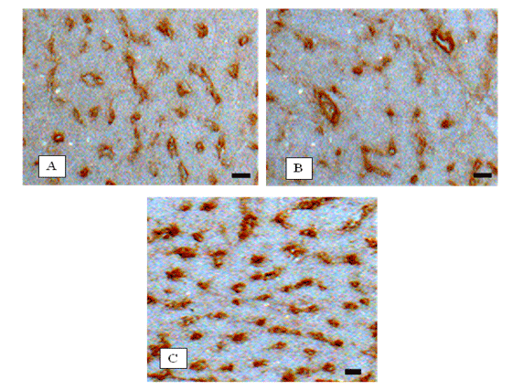
Figure 4: Representative image of the immunohistochemical CD 31 staining of LV capillaries, which appear brown in color: (A) MI-Ex (B) MI-Sed, and
(C) Sham. Capillaries are visualized using a light microscope at 20x magnification. The bar in the photomicrographs indicates 10 μm.
Discussion
In a rat model of severe MI, we demonstrated that exercise
training significantly increases capillary density in both the
LV and septum of the viable myocardium. Exercise-induced
angiogenesis such as that observed in our study may enhance
myocardial blood perfusion and improve cardiac functioning
in the infarcted heart. These data provide further insights into
the mechanisms underlying the improvement in morbidity and
mortality produced by exercise training in patients with MI.
Our results confirm previous evidence showing that low to moderate intensity treadmill exercise started late after MI beneficially impacts cardiac function and attenuates adverse remodeling [16, 23] . Contrarily, training programs consisting of higher exercise intensities, and started early after MI have reported detrimental effects on LV geometry and mortality in rats [13, 6]. The use of endurance swimming, which elicits higher mental and hemodynamic stresses compared with treadmill exercise, in addition to increased training frequency (90 min/d, 6 d/wk) strongly differentiates these previous exercise protocols from that used in the present investigation, and may account for the negative effects of exercise in HF animals observed by these authors [6].
In the present study, our results illustrated that MI-Ex capillary density in the septum and left ventricle was 1.4-fold higher (Figure 1) and 1.6-fold higher (Figure 3) than in MI-Sed (1220 ± 54 vs. 890 ± 43, and 950 ± 47 vs. 610 ± 44), suggesting that exercise promotes capillary growth in non-infarcted areas of severely decompensate hearts. Similarly, a study conducted by Leosco et al. reported that exercise induced a significant increase of capillary density in lateral border and remote zones of LV in HFEX hearts, but not in the LV anterior wall, which was largely involved in the infarcted area [15]. Although the pro-antigenic effect of exercise has previously been demonstrated in healthy swine hearts [22], data from the present study, in addition to the work of Leosco and colleagues, represents the only literature on exercise-dependent enhanced cardiac angiogenesis after MI. Our results showed that there was no significant difference in the ratio of heart weight to body weight regardless of the higher capillary density observed in the exercised trained LV. Although angiogenesis is contributed to hypertrophy of the heart [2]. Post-MI cardiac hypertrophy is mainly caused by angiotensin II mediated cell proliferation [8]. Our previous studies [21, 24] demonstrated that post-MI exercise training attenuated both plasma and cardiac tissue angiotensin II. This may explain why the heart weight to body weight ratio in the MI-EX group did not differ from their sedentary counterpart.
In conclusion, moderate intensity exercise training significantly increases capillary density in both the LV and septum of the viable myocardium. In this regard, exercise induced angiogenesis has the potential to impact numerous clinical conditions involving the ischemic and infarcted myocardium.
Our results confirm previous evidence showing that low to moderate intensity treadmill exercise started late after MI beneficially impacts cardiac function and attenuates adverse remodeling [16, 23] . Contrarily, training programs consisting of higher exercise intensities, and started early after MI have reported detrimental effects on LV geometry and mortality in rats [13, 6]. The use of endurance swimming, which elicits higher mental and hemodynamic stresses compared with treadmill exercise, in addition to increased training frequency (90 min/d, 6 d/wk) strongly differentiates these previous exercise protocols from that used in the present investigation, and may account for the negative effects of exercise in HF animals observed by these authors [6].
In the present study, our results illustrated that MI-Ex capillary density in the septum and left ventricle was 1.4-fold higher (Figure 1) and 1.6-fold higher (Figure 3) than in MI-Sed (1220 ± 54 vs. 890 ± 43, and 950 ± 47 vs. 610 ± 44), suggesting that exercise promotes capillary growth in non-infarcted areas of severely decompensate hearts. Similarly, a study conducted by Leosco et al. reported that exercise induced a significant increase of capillary density in lateral border and remote zones of LV in HFEX hearts, but not in the LV anterior wall, which was largely involved in the infarcted area [15]. Although the pro-antigenic effect of exercise has previously been demonstrated in healthy swine hearts [22], data from the present study, in addition to the work of Leosco and colleagues, represents the only literature on exercise-dependent enhanced cardiac angiogenesis after MI. Our results showed that there was no significant difference in the ratio of heart weight to body weight regardless of the higher capillary density observed in the exercised trained LV. Although angiogenesis is contributed to hypertrophy of the heart [2]. Post-MI cardiac hypertrophy is mainly caused by angiotensin II mediated cell proliferation [8]. Our previous studies [21, 24] demonstrated that post-MI exercise training attenuated both plasma and cardiac tissue angiotensin II. This may explain why the heart weight to body weight ratio in the MI-EX group did not differ from their sedentary counterpart.
In conclusion, moderate intensity exercise training significantly increases capillary density in both the LV and septum of the viable myocardium. In this regard, exercise induced angiogenesis has the potential to impact numerous clinical conditions involving the ischemic and infarcted myocardium.
Acknowledgement
This study was supported in part by a grant from the National
Heart, Lung, and Blood Institute (RO1-HL074273).
- Alhaddad IA, Hakim Ishrat, Siddiqi Faizi, Lagen back Edward, Mallavarapu Christopher, Nethala, Venkatram et al. Early exercise after experimental myocardial infarction: effect on left ventricular remodeling. Coron Artery Dis. 1998;9(6): 319-327.
- Anversa P, Beghi C, Kikkawa Y, Olivetti G. Myocardial infarction in rats. Infarct size, myocyte hypertrophy, and capillary growth. Circ Res. 1986;58(1):26-37.
- Bloor CM. Angiogenesis during exercise and training. Angiogenesis. 2005; 8(3): 263-271.
- Carlson CL, WW Winder. Liver AMP-activated protein kinase and acetyl-CoA carboxylase during and after exercise. J Appl Physiol. 1985;86(2):669-674.
- Egginton S. Invited review: activity-induced angiogenesis. Pflugers Arch.2009;457(5): 963-977.
- Gaudron P, Hu K, Schamberger R, Budin M, Walter B, Ertl G. Effect of endurance training early or late after coronary artery occlusion on left ventricular remodeling, hemodynamics, and survival in rats with chronic transmural myocardial infarction. Circulation. 1994;89(1):402-412.
- Giannuzzi P, Temporelli PL, Corrà U, Gattone M, Giordano A, Tavazzi L. Attenuation of unfavorable remodeling by exercise training in postinfarction patients with left ventricular dysfunction: results of the Exercise in Left Ventricular Dysfunction (ELVD) trial. Circulation. 1997;96(6): 1790-1797.
- Gray MO, Long CS, Kalinyak JE, Li HT, Karliner JS. Angiotensin II stimulates cardiac myocyte hypertrophy via paracrine release of TGF-beta 1 and endothelin-1 from fibroblasts. Cardiovasc Res. 1998;40(2):352-363.
- Gustafsson, TBodin K, Sylvén C, Gordon A, Tyni-Lenné R, Jansson E. Increased expression of VEGF following exercise training in patients with heart failure. Eur J Clin Invest. 2001;31(4):362-366.
- Iemitsu M, Maeda S, Jesmin S, Otsuki T, Miyauchi T. Exercise training improves aging-induced downregulation of VEGF angiogenic signaling cascade in hearts. Am J Physiol Heart Circ Physiol. 2006;291(3): H1290-1298.
- Jugdutt BI, Bogdan L Michorowski, C Tissa Kappagoda. Exercise training after anterior Q wave myocardial infarction: importance of regional left ventricular function and topography. J Am Coll Cardiol. 1988;12(2):362-372.DOI: 10.1016/0735-1097(88)90407-X
- Karam R, Healy BP, Wicker P. Coronary reserve is depressed in postmyocardial infarction reactive cardiac hypertrophy. Circulation. 1990;81(1):238-246.
- Kloner RA, JA Kloner . The effect of early exercise on myocardial infarct scar formation. Am Heart J. 1983;106(5-1): 1009-1013. doi.org/10.1016/0002-8703(83)90645-2
- Krichavsky MZ, DW Losordo. Prevention and recovery of hibernating myocardium by micro vascular repair. Circulation. 2011;124(9):998-1000. Doi.org/10.1161/CIRCULATIONAHA.111.047746
- Leosco, D Rengo G, Iaccarino G, Golino L, Marchese M, Fortunato F. Exercise promotes angiogenesis and improves beta-adrenergic receptor signalling in the post-ischaemic failing rat heart. Cardiovasc Res. 2008;78(2): 385-394.
- Musch TI, Moore RL, Leathers DJ, Bruno A, Zelis R. Endurance training in rats with chronic heart failure induced by myocardial infarction. Circulation. 1986;74(2): 431-441.
- Prior BM, Yang HT, Terjung RL. What makes vessels grow with exercise training?. J Appl Physiol. 2004;97(3): 1119-1128.
- Shiojima I, Kaori Sato, Yasuhiro Izumiya, Stephan Schiekofer, Masahiro Ito,Ronglih Liao, et al. Disruption of coordinated cardiac hypertrophy and angiogenesis contributes to the transition to heart failure." J Clin Invest. 2005;115(8): 2108-2118. Doi: 10.1172/JCI24682
- Sysa-Shah, P Yi Xu , Xin Guo, Frances Belmonte, Byunghak Kang, Djahida Bedja. Cardiac-Specific Over-Expression of Epidermal Growth Factor Receptor 2 (ErbB2) Induces Pro-Survival Pathways and Hypertrophic Cardiomyopathy in Mice." PLoS One. 2012;7(8):e42805.
- Veras-Silva AS, Mattos KC, Gava NS, Brum PC, Negrão CE, Krieger EM. Low-intensity exercise training decreases cardiac output and hypertension in spontaneously hypertensive rats. Am J Physiol. 1997;273(6 Pt 2): H2627-2631.
- Wan W, Anthony S PowersBS, Ji Li, John Q Zhang, Lisa Ji, John M.Erikson. Effect of post-myocardial infarction exercise training on the renin-angiotensin-aldosterone system and cardiac function. Am J Med Sci. 2007;334(4): 265-273. doi.org/10.1097
- White FC, Bloor CM, McKirnan MD, Carroll SM. Exercise training in swine promotes growth of arteriolar bed and capillary angiogenesis in heart. J Appl Physiol. 1998;85(3):1160-1168.
- Wisloff U, Jan P Loennechen, SusanCurrie, Godfrey L Smith, Øyvind Ellingsen. Aerobic exercise reduces cardiomyocyte hypertrophy and increases contractility, Ca2+ sensitivity and SERCA-2 in rat after myocardial infarction. Cardiovasc Res. 2002;54(1):162-174. doi.org/10.1016/S0008-6363(01)00565-X
- Xiaohua Xu, Wenhan Wan, Lisa Ji, Shunhua Lao, Anthony S Powers, Weiyan Zhao, John M Erikson. Exercise training combined with angiotensin II receptor blockade limits post-infarct ventricular remodelling in rats. Cardiovasc Res. 2008;78(3);523-532. Doi.org/10.1093/cvr/cvn028
- XiaohuaXu, Wenhan Wan, Anthony S Powers, JiLi, Lisa L Ji, ShunhuaLao et al. Effects of exercise training on cardiac function and myocardial remodeling in post myocardial infarction rats. J Mol Cell Cardiol. 2008;44(1):114-122. Doi.org/10.1016/j.yjmcc.2007.10.004

