


Methods: 184 bare metal stent (BMS) implanted patients were enrolled in this case-control study. According to coronary angiography results, all patients were divided into 2 groups: in-stent restenosis (ISR) group and non-ISR group. Serum Wnt5a levels were determined using enzyme-linked immune sorbent assay (ELISA).
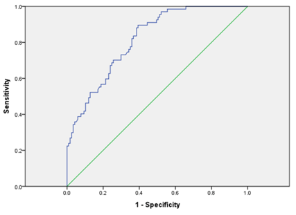
Results: Serum Wnt5a levels were higher in ISR group than those in non-ISR group, and were associated with the Gensini score. ISR rate was the highest in the upper tertile of Wnt5a. Multivariate logistic regression indicated that Wnt5a was an independent risk factor of ISR. For diagnosing ISR, the area under ROC curve was 0.817. The cutoff value of Wnt5a diagnosing ISR was 25.83ng/L, with the specificity of 60.68% and sensitivity of 89.55%.
Conclusion: Serum Wnt5a levels is associated with ISR in PCI patients with BMS implantation. Wnt5a is an independent risk factor of ISR, and may act as a biomarker for diagnosing ISR in BMS implanted patients.
Keywords: WNT5A; In-Stent Restenosis; Percutaneous coronary intervention; Bare metal stent.
ISR occurs in an average of 12 months in DES implanted patients and 6 months in bare-metal stents (BMS) implanted patients post-PCI [2]. The occurrence of ISR is a complex patho physiological process, and its mechanism is still not quite clear. Vascular injury post-PCI is an important initiating factor, which subsequently induced inflammatory response and vascular reparative process [3]. The vascular response to PCI is associated with cellular functions in many cell types, including endothelial cells and vascular smooth muscle cells (VSMCs) [4].
The Wnt signal transduction cascade plays an important role in many physiological and patho physiological processes. It’s a main regulator of development of mammals, and affect cell functions in many cell types [5]. Wnt proteins can activate two different pathways. The canonical Wnt signaling dependents on β-catenin, while non-canonical Wnt signaling is β-cateninindependent. Wnt5a, which can activate non-canonical Wnt signaling, is a member of Wnt family, and it has recently attracted attention of researchers. It is reported that Wnt5a may be implicated in the regulation of cell proliferation and migration in endothelial cells and VSMCs, which is critical for the occurrence of ISR [6, 7]. However, the clinical significance of Wnt5a in ISR is still not clear.
In this study, we aim to explore the association between serum Wnt5a level and ISR, and to assess the possibility of Wnt5a to be a predictor of ISR in patients with BMS implantation.
|
ISR(n=67) |
Non-ISR(n=117) |
P |
Sex (male %) |
47 (70.15%) |
73 (62.39%) |
0.288 |
Age |
62.28±10.76 |
60.50±8.77 |
0.223 |
BMI |
21.69±1.85 |
21.51±2.13 |
0.566 |
SBP |
132.79±19.68 |
131.20±15.60 |
0.546 |
DBP |
76.93±11.57 |
75.65±10.06 |
0.435 |
Gensini score |
66.75±12.28 |
33.11±10.96 |
<0.001 |
Risk factors of CAD |
|||
Smoking |
44 (65.67%) |
66 (56.41%) |
0.218 |
Hyperlipidemia |
38 (56.72%) |
53 (45.30%) |
0.136 |
Hypertension |
38 (56.72%) |
52 (44.44%) |
0.109 |
Culprit vessel |
|||
LM |
5 (7.6%) |
7 (5.98%) |
0.696 |
LAD |
55 (82.09%) |
89 (76.07%) |
0.341 |
LCX |
35 (52.24%) |
47 (40.17%) |
0.113 |
RCA |
49 (73.13%) |
86 (73.50%) |
0.956 |
Laboratory test |
|||
FBG(mmol/L) |
5.22±0.63 |
5.12±0.59 |
0.294 |
BUN(mmol/L) |
5.04±1.23 |
4.81±1.27 |
0.233 |
Cr(μmol/L) |
73.77±16.43 |
70.80±14.56 |
0.206 |
TC(mmol/L) |
4.14±0.97 |
4.18±1.09 |
0.818 |
TG(mmol/L) |
1.78±1.17 |
1.65±1.02 |
0.411 |
HDL(mmol/L) |
1.04±0.27 |
1.10±0.28 |
0.139 |
LDL(mmol/L) |
2.61±0.87 |
2.57±0.80 |
0.758 |
Wnt5a(ng/L) |
32.48±7.75 |
24.63±4.72 |
<0.001 |
IL-6(pg/ml) |
48.51±3.44 |
45.60±2.77 |
<0.001 |

Spearman correlation analysis showed that serum Wnt5a levels were associated with Gensini scores (r=0.580, P< 0.001).

ISR rate was the highest in tertile 3 (38/61) than those in tertile 2 (25/62) and tertile 3 (4/61).
|
OR (95% CI) |
P |
Univariate |
1.287(1.179,1.404) |
<0.001 |
Adjusted model1a |
1.287(1.177,1.408) |
<0.001 |
Adjusted model 2b |
1.284(1.174,1.405) |
<0.001 |
Adjusted model 3c |
1.256(1.129,1.397) |
<0.001 |
Model1a: Adjusted for sex, age and BMI
Model2b: Adjusted for model 1 and risk factors of CAD
Model3c: Adjusted for model 2 and TC, TG, LDL, HDL and IL-6
The mechanism of ISR is not fully elucidated. Kumatsu et al., investigated 11 stented coronary arteries in 11 patients. Histological findings indicated that inflammation and neointima proliferation is critical for the formation of ISR [11]. After PCI, the presence of stents can induce acute inflammation. Adhesion of acute inflammatory cells, including neutrophils and monocytes, is a character of early vascular response. Over weeks, the chronic inflammatory cells, including macrophages, replace acute inflammatory cells [4, 12]. Wnt5a is an important regulator of inflammation. Chiristman el al. reported that Wnt5a expressed in advanced atherosclerosis plaques, which is characterized by macrophage aggregation [13]. Recombinant Wnt5a could induce inflammatory cytokines production of macrophages via TLR4 dependent pathways, including IL-6 [14]. Our data showed that serum IL-6 levels, an important inflammatory cytokine, is higher in ISR group than those in non-ISR group. Multivariate logistic regression suggested that Wnt5a is independently associated with the presence of ISR, even adjusted for IL-6. This result indicates that Wnt5a may affect ISR by some other mechanism except for inflammation.
Neointima tissue proliferation is an important mechanism of ISR. This pathological process involves functional changes in various vascular wall cells. As the inner layer of arteries, endothelial cells play important roles in ISR process. Vascular endothelium integrity is an important prerequisite for protection of blood vessel [15]. However, PCI operation, including barotraumas and stimulation of stent, could damage vascular endothelium, which could induce the proliferation, migration and apoptosis of endothelial cells [16]. Wnt5a is reported to be a regulator of endothelial cells. Masckauchan el al., found that Wnt5a is expressed in human endothelial cells, and it could induce the proliferation and survival of endothelial cells. Endothelial cells migration was inhibited by reduced Wnt5a expression [17]. Kim et al. found that Wnt5a induced release of inflammatory cytokines, such as IL-6 and IL-8, via Wnt/Ca2+ signaling pathway. This result suggests that Wnt5a may act as an inflammatory mediator in endothelial cells [18].
Endothelial cell layer can inhibits VSMCs proliferation and intimal hyperplasia under normal circumstance, and VSMCs express high levels of contractile proteins. After vascular injury, phenotypic transformation may occur in VSMCs, which is characterized by increased level of proliferation, migration and extracellular matrix synthesis [19]. It is reported that the stable neointima contains 20% VSMCs [20], suggests that VSMCs play an important role in ISR. Pandey et al. found that Wnt5a mRNA was expressed in human VSMCs, as well as its receptors, such as Fzd1, Fzd2 and Fzd5 [21]. This result suggests that Wnt5a may be implicated in ISR via regulating VSMCs functions. Qin el al. treated VSMC with siRNA of Wnt5a, and found that total and free cholesterol content in VSMC exposed to oxLDL increased significantly, whereas recombinant Wnt5a treatment had the opposite effect [22]. This result suggests that Wnt5a could regulate cholesterol accumulation in VSMC. DirenZo el al. found that application of recombinant Wnt5a, Wnt2b, Wnt4 or Wnt9a stimulated VSMC proliferation via β-catenin dependent pathways [7]. However, it seems controversial that recombinant Wnt5a or over expression of Wnt5a cannot increase the proliferation of VSMC in another study [23]. Further study is needed to clarify Wnt5a functions in VSMC and underlying mechanisms.
Some limitations need to be noticed. First, this was a singlecenter study with a relatively small study population. It is necessary to confirm our conclusion in a large sample size study in the future. Second, as a case-control study, we cannot elucidate a causal relationship. Prospective and experimental studies are needed to fully elucidate the relationship between serum Wnt5a levels and ISR and its underlying mechanisms. Third, we did not measure serum Wnt5a levels over time. The dynamic changes in Wnt5a levels may help clarify the role of Wnt5a in the prognosis of ISR.
In conclusion, our data suggests that serum Wnt5a levels are associated with ISR in PCI patients with BMS implantation. Wnt5a is an independent risk factor of ISR, and it may act as a predict biomarker of ISR in BMS implanted patients. Our findings provide new insights into the clinical significance of the Wnt signaling pathway. Further study is needed to elucidate the relationship between Wnt5a and ISR and its underlying mechanisms.
- Piraino D, Cimino G, Buccheri D, Dendramis G, Andolina G, Cortese B. Recurrent in-stent restenosis, certainty of its origin, uncertainty about treatment. Int J Cardiol. 2017;230:91-96. doi.org/10.1016/j.ijcard.2016.12.073
- Lee MS, Banka G. In-stent Restenosis. Interv Cardiol Clin. 2016;5(2):211-220. doi: 10.1016/j.iccl.2015.12.006
- Alraies MC, Darmoch F, Tummala R, Waksman R. Diagnosis and management challenges of in-stent restenosis in coronary arteries. World J Cardiol. 2017;9(8):640-651. doi: 10.4330/wjc.v9.i8.640
- Pleva L, Kukla P, Hlinomaz O. Treatment of coronary in-stent restenosis: a systematic review. J Geriatr Cardiol. 2018;15(2):173-184. doi: 10.11909/j.issn.1671-5411.2018.02.007
- Nusse R, Clevers H. Wnt/beta-Catenin Signaling, Disease, and Emerging Therapeutic Modalities. Cell. 2017;169(6):985-999. doi: 10.1016/j.cell.2017.05.016
- Skaria T, Bachli E, Schoedon G. Wnt5A/Ryk signaling critically affects barrier function in human vascular endothelial cells. Cell Adh Migr. 2017;11(1):24-38. doi: 10.1080/19336918.2016.1178449
- DiRenzo DM, Chaudhary MA, Shi X, Franco SR, Zent J, Wang K, et al. A crosstalk between TGF-beta/Smad3 and Wnt/beta-catenin pathways promotes vascular smooth muscle cell proliferation. Cell Signal. 2016;28(5):498-505. doi: 10.1016/j.cellsig.2016.02.011
- Jimenez-Conde J, Biffi A, Rahman R, Kanakis A, Butler C, Sonni S, et al. Hyperlipidemia and reduced white matter hyperintensity volume in patients with ischemic stroke. Stroke. 2010;41(3):437-442. doi: 10.1161/STROKEAHA.109.563502
- Quan X, Ji Y, Zhang C, Guo X, Zhang Y, Jia S, et al. Circulating MiR-146a May be a Potential Biomarker of Coronary Heart Disease in Patients with Subclinical Hypothyroidism. Cell Physiol Biochem. 2018;45(1):226-236. doi: 10.1159/000486769
- Bhatt PM, Malgor R. Wnt5a: a player in the pathogenesis of atherosclerosis and other inflammatory disorders. Atherosclerosis. 2014;237(1):155-162. doi: 10.1016/j.atherosclerosis.2014.08.027
- Komatsu R, Ueda M, Naruko T, Kojima A, Becker AE. Neointimal tissue response at sites of coronary stenting in humans: macroscopic, histological, and immunohistochemical analyses. Circulation. 1998;98(3):224-233.
- Scott NA. Restenosis following implantation of bare metal coronary stents: pathophysiology and pathways involved in the vascular response to injury. Adv Drug Deliv Rev. 2006;58(3):358-376. DOI: 10.1016/j.addr.2006.01.015
- Christman MA, 2nd, Goetz DJ, Dickerson E, McCall KD, Lewis CJ, Benencia F, et al. Wnt5a is expressed in murine and human atherosclerotic lesions. Am J Physiol Heart Circ Physiol. 2008;294(6):H2864-2870. DOI: 10.1152/ajpheart.00982.2007
- Yu CH, Nguyen TT, Irvine KM, Sweet MJ, Frazer IH, Blumenthal A. Recombinant Wnt3a and Wnt5a elicit macrophage cytokine production and tolerization to microbial stimulation via Toll-like receptor 4. Eur J Immunol. 2014;44(5):1480-1490. doi: 10.1002/eji.201343959
- Flentje A, Kalsi R, Monahan TS. Small GTPases and Their Role in Vascular Disease. Int J Mol Sci. 2019;20(4). doi: 10.3390/ijms20040917
- Waksman R, Iantorno M. Refractory In-Stent Restenosis: Improving Outcomes by Standardizing Our Approach. Curr Cardiol Rep. 2018;20(12):140. doi: 10.1007/s11886-018-1076-6
- Masckauchan TN, Agalliu D, Vorontchikhina M, Ahn A, Parmalee NL, Li CM, et al. Wnt5a signaling induces proliferation and survival of endothelial cells in vitro and expression of MMP-1 and Tie-2. Mol Biol Cell. 2006;17(12):5163-5172. DOI: 10.1091/mbc.e06-04-0320
- Kim J, Kim J, Kim DW, Ha Y, Ihm MH, Kim H, et al. Wnt5a induces endothelial inflammation via beta-catenin-independent signaling. J Immunol. 2010;185(2):1274-1282. doi: 10.4049/jimmunol.1000181
- Kenagy RD. Biology of Restenosis and Targets for Intervention. In: Fitridge R, Thompson M, editors. Mechanisms of Vascular Disease: A Reference Book for Vascular Specialists. Adelaide (AU)2011.
- Clowes AW, Reidy MA, Clowes MM. Mechanisms of stenosis after arterial injury. Lab Invest. 1983;49(2):208-215.
- Pandey S, Chandravati. Wnt signaling cascade in restenosis: a potential therapeutic target of public health relevance in a North American cohort of Nebraska State. Mol Biol Rep. 2014;41(7):4549-4554. doi: 10.1007/s11033-014-3325-0
- Qin L, Hu R, Zhu N, Yao HL, Lei XY, Li SX, et al. The novel role and underlying mechanism of Wnt5a in regulating cellular cholesterol accumulation. Clin Exp Pharmacol Physiol. 2014;41(9):671-678. doi: 10.1111/1440-1681.12258
- Qin L, Liu C, Ni YG, Shi YN, Ao BX, Liu Z, et al. OS 02-07 WNT5A/ROR2 SUPPRESSES THE PROLIFERATION OF SMOOTH MUSCLE CELL VIA PKC SIGNALING PATHWAY. Journal of Hypertension. 2016;34:e49. doi: 10.1097/01.hjh.0000499982.63412.6a

