


Methods: This study included 25 patients with diabetes but no severe organ damage (type 1 diabetes [T1D]: 12 cases, type 2 diabetes [T2D]: 13 cases). The patients were evaluated to determine their RHI and its relation with continuous glucose monitoring (CGM) parameters, glycated hemoglobin (HbA1c), glycoalbumin (GA), high-density lipoprotein-cholesterol (HDL-C), triglycerides (TG), and tumor necrosis factor (TNF-α). Various other clinical and laboratory parameters were analyzed for associations with RHI.
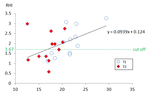
Results: Mean ages were 40±12 years in the T1D group and 54±13 years in the T2D group. Mean HbA1c levels were 7.6±1.0% in the T1D group and 7.0 ± 0.9% in the T2D group. RHI was positively correlated with GA among all cases (p=0.02) and among T1D cases (p=0.011). Among T2D cases, RHI was positively correlated with HDL-C (p=0.034) and negatively correlated with TG (p=0.03) and TNF-α (p=0.047). In the multivariate analysis, RHI was independently correlated with GA among all cases (β=0.471, p=0.02) and among T1D cases (β=0.727, p=0.011). RHI was also independently correlated with HDL-C among T2D cases (β=0.586, p=0.045). Among T1D cases, GA was negatively correlated with hypoglycemia frequency (p=0.005).
Conclusions: Endothelial dysfunction may be related to hypoglycemia in T1D and to HDL-C in T2D
Keywords: Vascular Endothelial Function; RH-PAT Index; Type 1 Diabetes Mellitus; Type 2 Diabetes Mellitus; Continuous Glucose Monitoring; Hypoglycemia;
Recent research has led to the introduction of a new method for measuring peripheral arterial hyperemia (reactive hyperemia peripheral arterial tonometry [RH-PAT]), which has low interexaminer variation and good reproducibility [4, 5]. The RH-PAT technique is used to calculate an index of vascular endothelial function (RH-PAT, RHI), which is correlated with various cardiovascular risk factors, such as body mass index (BMI), total cholesterol, high-density lipoprotein cholesterol (HDL-C), diabetes, and smoking [6]. Previous reports have also indicated that other indices of vascular endothelial function, such as FMD, are negatively correlated with the magnitude of blood glucose fluctuations and improve when blood glucose fluctuations are minimized [7–9]. However, few reports have addressed the relationship between RHI and daily blood glucose fluctuations [10, 11]. Therefore, the present study used RHI to evaluate vascular endothelial functions among patients with diabetes and used continuous glucose monitoring (CGM) to examine blood glucose parameters. These results were used to examine the associations between RHI and multiple arteriosclerosis-related factors, such as tumor necrosis factor-alpha (TNF-α), interleukin-6 (IL-6), and advanced glycation end products (AGEs) according to the type of diabetes.
All patients with T1D were female and were treated using multiple daily insulin injections. Among patients with T2D, 1 patient was receiving insulin injections, 5 patients were receiving insulin injections and oral hypoglycemic agents, 6 patients were receiving only medication, and 2 patients were being treated using diet therapy. None of patients were used insulin pump, All patients underwent evaluations to determine their RHI, and 3 patients were excluded because of device errors, and data from 25 patients (12 with T1D and 13 with T2D) were included in the study. Patients were considered excluded from participation if they had active infections, severe liver/kidney diseases, eating disorders, or psychiatric disorders. All patients provided written informed consent prior to participation, and our study protocol was approved by the ethics committee of Tokyo Women’s Medical University (131009; January 22, 2014).
Daily insulin dose (U/kg/day) was obtained from the patients’ medical records. Fluctuations in blood glucose were evaluated based on the means of daily differences (MODD) [13], mean amplitude of glycemic excursions (MAGE) [14], J-index [15], and M-value [16]. MODD was defined as the average value for differences in blood glucose at the same time during two consecutive days. MAGE was defined as the average amplitude of blood glucose fluctuations that exceeded 1 standard deviation for the 24-h average blood glucose value from CGM. The J-index was calculated as 0.324 × (mean blood glucose level + standard deviation)2. The M-value was defined as the average deviation of measured blood glucose levels from the basic value of 100 mg/ dL.
T1D |
T2D |
P |
|
n |
12 |
13 |
|
Sex (male/female) |
0/12 |
5/8 |
|
Age (years) |
41 ± 13 |
53 ± 13 |
<0.05 |
Duration of diabetes (years) |
17.3 ± 11.0 |
12.4 ± 9.2 |
0.444 |
BMI (kg/m2) |
24.6 ± 4.0 |
27.0 ± 4.0 |
0.162 |
Abdominal circumference (cm) |
80.6 ± 10.4 |
89.3 ± 9.0 |
<0.05 |
HbA1c (%) |
7.5 ± 1.0 |
7.0 ± 0.9 |
0.232 |
sBP (mmHg) |
123.0 ± 21.0 |
135.5 ± 10.5 |
0.073 |
dBP (mmHg) |
76.5 ± 10.4 |
78.5 ± 8.4 |
0.606 |
HR (bpm) |
81.7 ± 13.8 |
74.6 ± 15.0 |
0.243 |
TG (mg/dl) ※ |
103.7 ± 77.5 |
88.7 ± 40.3 |
0.689 |
LDL-C (mg/dl) |
109.1 ± 31.1 |
108.8 ± 25.0 |
0.978 |
HDL-C (mg/dl) |
79.5 ± 15.4 |
60.0 ± 14.8 |
<0.05 |
eGFR (ml/min) ※ |
87.5 ± 22.9 |
83.2 ± 24.3 |
0.47 |
ACR (mg/gCr) ※ |
30.2 ± 83.4 |
5.3 ± 4.4 |
0.769 |
UA (mg/dl) |
3.8 ± 1.0 |
5.5 ± 1.3 |
<0.05 |
RHI |
2.08 ± 0.74 |
1.83 ± 0.70 |
0.194 |
Student’s t -test |
※Mann-Whitney U test |
Variable |
R |
P |
Age(years) |
-0.065 |
0.757 |
Duration(years) |
0.137 |
0.513 |
BMI(kg/m2) |
-0.325 |
0.112 |
HbA1c(%) |
0.262 |
0.206 |
GA(%) |
0.471 |
0.02 |
LDL-C(mg/dl) |
0.063 |
0.764 |
HDL-C(mg/dl) |
0.337 |
0.099 |
eGFR(ml/min) |
-0.062 |
0.768 |
High-sensitivity CRP(ng/ml) |
-0.352 |
0.099 |
Pentosidine (μg/ml) |
-0.225 |
0.302 |
TNF-α(pg/ml) |
-0.046 |
0.835 |
MODD |
0.201 |
0.347 |
MAGE |
0.066 |
0.755 |
J-INDEX |
0.323 |
0.116 |
M-value※ |
0.293 |
0.155 |
CGM mean blood glucose(mg/dl) |
0.166 |
0.428 |
Frequency of hypoglycemia below 70mg/dl (/day) |
-0.114 |
0.586 |
hypo: hypoglycemia |
Pearson’s correlation coefficient |
※Spearman |

Variable |
R |
P |
Age (years) |
-0.24 |
0.453 |
Duration of diabetes (years) |
-0.277 |
0.383 |
BMI (kg/m2) |
-0.406 |
0.191 |
FBG (mg/dl) |
-0.414 |
0.181 |
HbA1c (%) |
0.295 |
0.351 |
GA (%) |
0.727 |
0.011 |
TG (mg/dl)※ |
0.291 |
0.359 |
LDL-C (mg/dl) |
0.090 |
0.781 |
HDL-C (mg/dl) |
-0.008 |
0.98 |
eGFR (ml/min) |
0.200 |
0.532 |
High-sensitivity CRP (ng/ml)※ |
-0.373 |
0.259 |
Pentosidine (μg/ml)※ |
-0.582 |
0.06 |
TNF-α (pg/ml) |
0.314 |
0.347 |
MODD |
0.298 |
0.347 |
MAGE |
0.138 |
0.669 |
J-INDEX |
0.445 |
0.148 |
CGM mean blood glucose(mg/dl) |
0.53 |
0.077 |
Frequency of hypo <70mg/dl(/day) |
-0.522 |
0.082 |
hypo: hypoglycemia |
Pearson’s correlation coefficient |
※Spearman |
Variable |
R |
P |
Age (years) |
0.256 |
0.398 |
Duration of diabetes (years) |
0.531 |
0.062 |
BMI (kg/㎡) |
-0.183 |
0.55 |
FBG (mg/dl) |
0.047 |
0.88 |
HbA1c (%) |
0.156 |
0.611 |
GA (%) |
0.171 |
0.576 |
TG (mg/dl)※ |
-0.601 |
0.03 |
LDL-C (mg/dl) |
0.031 |
0.919 |
HDL-C (mg/dl) |
0.588 |
0.034 |
eGFR (ml/min) |
-0.337 |
0.261 |
High-sensitivity CRP (ng/ml)※ |
-0.49 |
0.106 |
Pentosidine (μg/ml)※ |
-0.217 |
0.498 |
TNF-α (pg/ml) |
-0.583 |
0.047 |
MODD |
-0.004 |
0.991 |
MAGE |
-0.27 |
0.373 |
J-INDEX |
-0.27 |
0.372 |
CGM mean blood glucose(mg/dl) |
-0.051 |
0.868 |
Frequency of hypo < 70mg/dl(/day) |
0.17 |
0.58 |
hypo: hypoglycemia |
Pearson’s correlation coefficient |
※Spearman |

Among 4,352 patients from various cohorts of the Framingham study (55 ± 16 years old, 51% female), RHI was significantly associated with age, systolic blood pressure, heart rate, BMI, total cholesterol/HDL-C ratio, diabetes, and smoking [18]. Furthermore, among 106 Japanese patients with T2D, RHI was significantly associated with the ankle-brachial index, carotid IMT, and statin use [19]. In the present study, RHI was not correlated with GA among patients with T2D, although RHI was correlated with lipid profile factors, TNF-α, and HDL-C. These results indicate that RHI is correlated with arteriosclerotic factors and supports the results of the Framingham study [18] and those by Ueda et al. [19].
A previous study indicated that patients with T1D have remarkably lower RHI values than non-diabetic patients [20]. In addition, patients with T1D and endothelial dysfunction based on their RHI value tend to have high HbA1c levels [21]. A prospective study of 73 young patients with T1D suggested that RHI improved with better control of HbA1c [22], although this relationship was not observed in the present study.
The present study revealed that among patients with averaged 16-year durations of T1D, RHI was correlated with GA. In addition, GA was negatively correlated with the frequency of hypoglycemia (< 70 mg/dL; r = –0.727, p = 0.011), which suggests that RHI may decrease with hypoglycemia. This possibility is also supported by the fact that RHI may be reduced during vasoconstriction caused by sympathetic nerve stimulation. Furthermore, among patients with T1D, FMD values were found to be negatively correlated with two indicators of hypoglycemia (glycemic risk assessment diabetes equation [GRADE] and low blood glucose index [LBGI]) [23]. Moreover, patients with T1D and frequent severe hypoglycemic episodes exhibited higher levels of plaque in the carotid and femoral arteries, as well as notably increased serum markers of inflammation [24]. Although it remains unclear how hypoglycemia can influence changes in vascular function, hypoglycemia is known to stimulate the sympathetic nervous system to increase heart rate, cardiac output, myocardial contraction, systolic blood pressure, plasminogen activation inhibitor, and inflammatory reactions (e.g., through platelet activity, P-selectin, and high-sensitivity CRP) [25, 26]. Thus, patients with diabetes who experience constant oxidative stress during hypoglycemia may experience compensatory physical changes, hematological changes, and an increased inflammatory response.
The onset or development of diabetic complications may be mediated by AGEs, which bind to receptors for AGE (RAGE) on almost all cells and activate intracellular signals. In this context, the AGE–RAGE system is a trigger of oxidative stress and is related to inflammation and apoptosis [27]. Furthermore, the accumulation of AGEs causes arteriosclerotic disease, and is involved in the development of microangiopathy and macroangiopathy in patients with diabetes. Although AGEs are through to decrease NO synthase expression in the vascular endothelium and subsequently decrease FMD-measured vascular endothelial function [28], no reports have suggested that AGEs may be related to RHI. The present study also failed to detect an association between RHI and serum pentosidine (one of the AGEs) among patients with T1D or T2D. However, further studies are needed to address this issue, since the present study only examined a small sample of young patients.
The present study demonstrated that it is possible to examine the relation between RHI and arteriosclerosis-related factors or CGM parameters. Another possible strength of the present study is that a single examiner performed all RHI measurements, which indicates that the measurement error may be very small. However, the present study is limited by the small sample size (28 patients) and the fact that 23 of the 28 patients were women.
- Schächinger V, Britten MB, Zeiher AM. Prognostic impact of coronary vasodilator dysfunction on adverse long-term outcome of coronary heart disease. Circulation. 2000;101(16):1899–1906.
- Corretti MC, Anderson TJ, Benjamin EJ, Celermajer D, Charbonneau F, Creager MA, et al. Guidelines for the ultrasound assessment of endothelial-dependent flow-mediated vasodilation of the brachial artery: a report of the International Brachial Artery Reactivity Task Force. J Am Coll Cardiol. 2002;39(2):257–265.
- Thijssen DH, Black MA, Pyke KE, Padilla J, Atkinson G, Harris RA, et al. Assessment of flow-mediated dilation in humans: a methodological and physiological guideline. Am J Physiol Heart Circ Physiol. 2011;300(1):H2–12. doi: 10.1152/ajpheart.00471.2010
- Bonetti PO, Barsness GW, Keelan PC, Schnell TI, Pumper GM, Kuvin JT et al. Enhanced external counterpulsation improves endothelial function in patients with symptomatic coronary artery disease. J Am Coll Cardiol. 2003;41(10):1761–1768.
- Kuvin JT, Patel AR, Sliney KA, et al. Assessment of peripheral vascular endothelial function with finger pulse wave amplitude. Am Heart J. 2003;146(1):168–174. Doi: 10.1016/S0002-8703(03)00094-2
- Hamburg NM, Keyes MJ, Larson MG, Pandian NG, Sheffy J, Schnall RP,et al. Cross-sectional relations of digital vascular function to cardiovascular risk factors in The Framingham Heart Study. Circulation. 2008;117:2467–2474. Doi: 10.1016/S0002-8703(03)00094-2
- Maiorino MI, Casciano O, Della Volpe E, Bellastella G, Giugliano D, Esposito K. et al. Reducing glucose variability with continuous subcutaneous insulin infusion increases endothelial progenitor cells in type 1 diabetes: an observational study. Endocrine. 2016;52(2):244–252. Doi: 10.1007/s12020-015-0686-7
- Harrington J, Peña AS, Wilson L, et al. Vascular function and glucose variability improve transiently following initiation of continuous subcutaneous insulin infusion in children with type 1 diabetes. Pediatr Diabetes. 2013;14(7):504–511. Doi: 10.1111/pedi.12050
- Jamiołkowska M, Jamiołkowska I, Łuczyński W, et al. Impact of Real-Time Continuous Glucose Monitoring Use on Glucose Variability and Endothelial Function in Adolescents with Type 1 Diabetes: New Technology-New Possibility to Decrease Cardiovascular Risk? J Diabetes Res. 2016;2016:4385312. Doi: 10.1155/2016/4385312
- Torimoto K, Okada Y, Mori H, Otsuka T, Kawaguchi M, Matsuda M, et al. Effects of exenatide on postprandial vascular endothelial dysfunction in type 2 diabetes mellitus. Cardiovasc Diabetol. 2015;14:25. Doi: 10.1186/s12933-015-0188-1
- Torimoto K, Okada Y, Mori H, Tanaka Y. Relationship between fluctuations in glucose levels measured by continuous glucose monitoring and vascular endothelial dysfunction in type 2 diabetes mellitus. Cardiovasc Diabetol. 2013;12:1. Doi: 10.1186/1475-2840-12-1
- Matsuo S, Imai E, Horio M, et al. Revised equations for estimated GFR from serum creatinine in Japan. Am J Kidney Dis. 2009;53(6):982–992. Doi: 10.1053/j.ajkd.2008.12.034
- Molnar GD, Taylor WF, Ho MM. Day-to-day variation of continuously monitored glycaemia: a further measure of diabetic instability. Diabetologia. 1972;8(5):342–348.
- Service FJ, Molnar GD, Rosevear JW, Ackerman E, Gatewood LC, Taylor WF. Mean amplitude of glycemic excursions, a measure of diabetic instability. Diabetes. 1970;19(9):644–655.
- Wójcicki JM. “J”-index. A new proposition of the assessment of current glucose control in diabetic patients. Horm Metab Res. 1995;27(1):41–42.
- Schlichtkrull J, Munck O, Jersild M. The M-Value, an Index of Blood-sugar Control in Diabetics. Acta Medica Scandinavica. 1965;177:95–102.
- Bonetti PO, Pumper GM, Higano ST, Holmes DR Jr, Kuvin JT, Lerman A, et al. Noninvasive identification of patients with early coronary atherosclerosis by assessment of digital reactive hyperemia. J Am Coll Cardiol. 2004;44(11):2137–2141. Doi: 10.1016/j.jacc.2004.08.062
- Hamburg NM, Palmisano J, Larson MG, et al. Relation of brachial and digital measures of vascular function in the community: the Framingham Heart Study Hypertension. 2011;57(3):390–396. Doi: 10.1161/HYPERTENSIONAHA.110.160812
- Ueda M, Takabe M, Sawada T, et al. Comparison Between 2 Types of Vascular Endothelial Function Test (Flow-Mediated Dilatation vs. Reactive Hyperemia Index) in Patients With Type 2 Diabetes Mellitus. J Jap Diabetes Society. 2017;60(5):370–377. Doi: 10.11213/tonyobyo.60.370
- Pareyn A, Allegaert K, Asscherickx W, et al. Impaired endothelial function in female adolescents with type 1 diabetes measured by peripheral artery tonometry. Eur J Pediatr. 2013;172(8):1017–1022. Doi: 10.1007/s00431-013-1988-5
- Palombo C, Kozakova M, Morizzo C, Gnesi L, Chiara Barsotti M, Spontoni M, et al. Circulating endothelial progenitor cells and large artery structure and function in young subjects with uncomplicated type 1 diabetes. Cardiovasc Diabetol. 2011;10:88–97. Doi: 10.1186/1475-2840-10-88
- Scaramuzza AE, Redaelli F, Giani E, Macedoni M, Giudici V, Gazzarri A, et al. Adolescents and young adults with type 1 diabetes display a high prevalence of endothelial dysfunction. Acta Pediatrica. 2015;104(2):192–197. Doi: 10.1111/apa.12877
- Peña AS, Couper JJ, Harrington J, et al. Hypoglycemia, but Not Glucose Variability, Relates to Vascular Function in Children with Type 1 Diabetes. Diabetes Technol Ther. 2012;14(6):457–462. Doi: 10.1089/dia.2011.0229
- Giménez M, Gilabert R, Monteagudo J, Anna Alonso, Roser Casamitjana, Ignacio Conget. Repeated Episodes of Hypoglycemia as a Potential Aggravating Factor for Preclinical Atherosclerosis in Subjects With Type 1 Diabetes. Diabetes Care. 2011;34(1):198–203. Doi: 10.2337/dc10-1371
- Gogitidze JN, Hedrington MS, Briscoe VJ, Tate DB, Ertl AC, Davis SN. Effects of acute hypoglycemia on inflammatory and pro-atherothrombotic biomarkers in individuals with type 1 diabetes and healthy individuals. Diabetes Care. 2010;33(7):1529–1535. Doi: 10.2337/dc09-0354
- Wright RJ, Newby DE, Stirling D, Ludlam CA, Macdonald IA, Frier BM. Effects of acute insulin-induced hypoglycemia on indices of inflammation: putative mechanism for aggravating vascular disease in diabetes. Diabetes Care. 2010;33(7):1591–1597. Doi: 10.2337/dc10-0013
- Stirban A, Gawlowski T, Roden M, et al. Vascular effects of advanced glycation end products: Clinical effects and molecular mechanisms. Mol Metab. 2013;3(2):94–108. Doi: 10.1016/j.molmet.2013.11.006
- Xiaomei Ren, Ligun Ren, Qin Wei, Shao H, Chen L, Liu N. Advanced glycation end-products decreases expression of endothelial nitric oxide synthase through oxidative stress in human coronary artery endothelial cells. Cardiovasc Diabetol. 2017;16(1):52. Doi: 10.1186/s12933-017-0531-9

