


2Department of Internal Medicine and Specialties; Faculty of medicine and biomedical sciences, University of Yaoundé I
3Urology unit Yaoundé Central Hospital
The occurrence of an adrenal insufficiency post adrenalectomy usually attests of the success of the operative act. After the adrenalectomy, we could expect a compensatory secretion by the controlateral adrenal gland. However, the integrity of the residual function of the remaining gland depends on the initial secretory character of the mass and on its length. The aim of this study was to evaluate the residual function of the controlateral gland after a unilateral adrenalectomy.
Method:
This was a 13-month prospective study in Yaoundé Central Hospital. We included in the study, all patients who underwent an adrenalectomy. The adrenal function was assessed in preoperative by the measurement of 8am plasmatic cortisol after a dexamethasone suppression test, while in postoperative periods it was the measure of the baseline plasmatic cortisol at 8am. The latter was possibly completed with a stimulation test with Synacthene 0.25μg. The association between the variables was searched using the Fischer test. A significance threshold of 0.05 was adopted.
Results:
Seven patients (4 women and 3 men) underwent surgery indicated for an adrenal mass. Median age was 44,7 years [17 – 69 years]. The discovery mode was mainly weight gain (28.6 %) or unexplained weight loss (28.6 %). The median delay to diagnosis was 8months [8 days – 24 months]. In the preoperative period, the median cortisol level after dexamethasone suppression test was 329.9 ng/ml, amongst which 2/7 patients had hypercortisolism. The median values of normetanephrines and metanephrines were 7nmol/L and 71.4 nmol/L, respectively. Pathology described: corticosurrenalomas (2), adrenal adenomas (2), pheochromocytomas (2) and adrenalitis (1). In early postoperative, the median 8 h cortisol was 45.5 ng/ml [34.5-167.1ng/ml]. In late postoperative, the median cortisol value was 95 ng/ml, and strictly normal in 3/6 patients. There was no association between recovery of residual adrenal function and age, tumor size, initial preoperative cortisol value, treatment received preoperatively, and postoperative complications.
Conclusion
Adrenal insufficiency persists in almost half of the patients in late postoperative. Thus, hydrocortisone supplementation should be maintained as long as possible.
We conducted a prospective open-label cohort study during a 13-month period (i.e., July 2020 to July 2021) at the Endocrinology Department of HCY. The source population consisted of patients operated upon for adrenal mass at the Central Hospital of Yaoundé. The sample was consecutive and exhaustive.
• Data collection:
We identified patients operated and followed up at the endocrinology and urology departments during the defined study period from the operating room register and during endocrinology consultations. Then, we collected the data from a pre-established data collection form. The form provided information on sociodemographic data, personal and family history, preoperative
clinical examination, paraclinical examinations: biological and CT scan, diagnosis after anatomopathological analysis, as well as the treatment received pre and postoperatively. Missing data were completed after questioning and re-evaluation of these patients. Simultaneous recruitment of newly diagnosed patients was also performed from the pre- to the postoperative phase. Clinical monitoring of all patients was performed, describing the clinical and paraclinical evolution of patients in the immediate, early and late postoperative phases. Patients were followed up every three months.
Clinically, at each visit we looked for functional signs of adrenal insufficiency, mainly asthenia, digestive disorders and signs related to hypotension. Then the patients were subjected to a complete physical examination after which they were systematically sampled for biological analyses according to the previously defined schedule.
• Assessment of adrenal function (4):
Adrenal function was assessed by measuring plasma cortisol. In the pre-operative period, we systematically looked for hypercortisolism by measuring the midnight cortisol followed by a low-dose overnight dexamethasone suppression test. The latter consisted in administering 1 mg of dexamethasone, which the patient had to take at 11 pm, followed by the dosage of 8am cortisol after suppression the next day. In the post-operative, the baseline 8am cortisol test was measured. We measured cortisol at least twice: early postoperative (0 to 3 months) and late postoperative (> 3 months).
The cortisol measurement was completed by a stimulation test with Synacthen® when the baseline cortisol was normal. This was to ensure that there is a total recovery of the adrenal function, The Synacthen® stimulation test consisted of taking a baseline cortisol sample at 8:00 am. Then an IV injection of ACTH analogue (0.25 mg Synacthen®) was given. Then samples were taken at T0 and T60 min for plasma cortisol determinations.
• Interpretation: (8)
Cortisol at 8:00 am after dexamethasone suppression test >18ng/ml (50nmol/l): hypercortisolism. 8am baseline cortisol: 50 -180 ng/ml (138 - 500 nmol/L): Normal Cortisol at T60 min after Synacthen test <18 ng/ml: Adrenal insufficiency
• Statistical analysis:
Data were entered and analyzed using Statistical Package for the Social Sciences (SPSS) software version 26.0. Charts and tables were prepared using Word and Excel 2016 software. Quantitative variables were expressed either as mean ± standard deviation or as median and interquartile range. Qualitative variables were expressed as headcount and percentage. The association between the variables was investigated using the Fischer test; the measure of association used was the relative risk. The statistical significance level was set at 0.05; with a 95% confidence interval.
During the study period, the endocrinology department of HCY recorded 781 endocrinology consultations apart from diabetes, and 17 (2%) of these were adrenal tumor-related pathologies. Of the 8 cases with adrenal mass, 7 were operated on and followed up during the predefined period. Our series of patients was predominantly female, with a sex ratio of 1.3. Most of them were older than 40 years, with a median age at diagnosis around 44 years, and extremes between 17 - 69 years.
2. Clinical data:
a. Mode of discovery:
The main mode of discovery of adrenal masses was: unexplained weight loss and weight gain. In addition, there was resistant hypertension, thoraco-abdominal pain and difficulty in urination.
The median duration of progression from symptom onset to diagnosis was 240 days (eight months), with a minimum of eight days and a maximum of 720 days (24 months). No patient had a personal or family history of adrenal tumors, or other tumors in favor of a predisposition syndrome to them.
d. Preoperative treatment
One of the two patients with severe Cushing’s syndrome was put on high dose Ketoconazole. And patients with hypertension were put on antihypertensive drugs.
e. Operative indications, type of procedure:
The operative indications were secreting masses 4 and 3 incidentalomas larger than 4 cm. All patients underwent open surgery by the same team, using a subcutaneous approach.
3. Paraclinical data:
a. Preoperative Hormonology. The hormonal workup was performed in the preoperative period, given the clinical presentation of the patients as described in the table 1. The median value of the preoperative cortisol post suppression test was 329.9ng/ml (122nmol/ml) and the midnight cortisol was 387.4ng/ml (143nmo/ml). Other hormones were not sufficiently reporte.
Table 1:
b. Description of the scannographic data
Adrenal masses diagnosed preoperatively were predominantly located on the right side, measuring more than 4 cm, with a median size of 5 cm [3.8 - 8cm]. The spontaneous density was > 10 HU. The median absolute washout was 50% [36 - 58.7] and the mean relative washout was 54 ± 19.79%. These masses showed no necrosis, calcification, hemorrhage, or infiltration Table 2.
c. Postoperative adrenal function assessment:
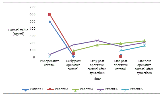
• In early postoperative (0 to 3 months): The median 8am cortisol value in early postoperative was 45.51 ng/ml, with a minimum of 34.50 ng/ml and a maximum of 167.10 ng/ml. Two of these patients had normal 8am cortisol values, which required a Synacthen test with responses of 165 and 226.20 ng/L, respectively.
• Late postoperative adrenal function (>3 months):
Regarding the late postoperative 8am cortisol, the one performed every three months from the 3rd postoperative month, six out of seven results (85.71%) were informed. A median value of 95 ng/L was obtained. This means a minimum of 7.00 ng/L and a maximum of 187.70 ng/L as baseline cortisol.
At the end of the study, three out of six patients (50%) had adrenal function within the normal range. And the other three remained in adrenal insufficiency (Figure1; Table3). d. Anatomo-pathological data:
Hormone |
Frequency (%) |
Médiane (IQ) |
Minimum |
Maximum |
Midnight cortisol (ng/ml) |
3 (42.9) |
387.3 (34.5 - //) |
34 .5 |
387.3 |
Cortisol DXM suppression test |
4 (57.1) |
329.9 (50 – 562.7) |
10.0 |
587.7 |
ACTH at midnight (pg/L) |
2 (28.6) |
- |
- |
< 1 |
24h UFC |
1 (14.3) |
- |
- |
23 |
11-DOC |
1 (14.3) |
- |
- |
0.1 |
Aldosterone (pmol/L) |
1 (14.3) |
- |
- |
390 |
DHEAS |
1 (14.3) |
- |
- |
3.7 |
Normetanephrine |
4 (57.14) |
7 (0.3 – 48.3) |
0.3 |
59.9 |
Metanephrine (nmol/L) |
5 (71.4) |
0.5 (0.2 – 8.8) |
0.1 |
28 |
Characteristics of masses |
Effective |
Percentage |
Localisation |
||
Right |
4 |
57,1 |
Left |
2 |
28,6 |
Bilatéral |
1 |
14,3 |
Height |
||
≥ 4 cm |
5 |
71,4 |
< 4 cm |
2 |
28,6 |
Spontaneous Density |
|
|
> 10 HU |
5 |
71,4 |
Patient |
8am Cortisol post DXM suppression test (ng/ml) |
8am baseline cortisol early post operative (0-3mois) (ng/ml) |
Cortisol T60 post Synacthene post-opérative (0-3mois) (ng/ml) |
8 am baseline cortisol in late post operative (>3mois) (ng/ml) |
Cortisol T60 post- Synacthene in late post operative (>3mois) (ng/ml) |
Patient 1 |
487.9 |
6.6 |
- |
28.5 |
- |
Patient 2 |
587.7 |
45.5 |
- |
7 |
- |
Patient 3 |
- |
84 |
165 |
189.7 |
223.6 |
Patient 4 |
34.5 |
167.1 |
226.2 |
143 |
199 |
Patient 5 |
10 |
- |
- |
95 |
163 |
Patient 6 |
- |
14 |
- |
- |
- |
Patient 7 |
- |
- |
- |
22.9 |
- |
4. Postoperative complications:
Complications that occurred postoperatively were predominantly infections of the surgical wound (6/7), followed by acute adrenal insufficiency in 5 patients. In addition, hydroelectrolytic disorders marked by hypokalemia and the occurrence of hypertension were also observed.
4. Bivariate analyses of factors influencing adrenal function postoperatively:
We found no association between recovery of residual adrenal function and age, tumor size, initial preoperative 8am cortisol value,
Variable |
Adrenalfunction |
RR |
IC à 95% |
p-value |
|
Altered (%) |
Normal (%) |
|
|
|
|
|
|
|
|
|
|
Age |
|
|
|
|
|
< 40 years |
2(100) |
0 |
- |
- |
|
≥ 40years |
1(25) |
3(75) |
4 |
0,73-21,83 |
0,4 |
Size of tumor |
|
|
|
|
|
≥ 4 cm |
1(25) |
3(75) |
0,25 |
0,046-1,36 |
0,4 |
<4 cm |
2(100) |
0 |
- |
- |
|
Post operative complications |
2(50) |
2(50) |
0,5 |
0,18-1,33 |
1 |
Pre operative Cortisol |
|
|
|
|
|
Hypersecretion |
2(100) |
0 |
- |
- |
|
Hyposecretion |
0 |
1(100) |
- |
- |
|
Comorbidities |
1(50) |
1(50) |
1 |
0,18-5,46 |
1 |
Previous traitement |
2(50) |
2(50) |
1 |
0,18-5,46 |
1 |
The results show that in the hormonal field, many data were missing due to the lack of financial means. This obliged the prescribers to contextualize the request for examinations by orienting them according to the clinical presentation. However, concerning the cortisol values, preoperatively, after the overnight DXM suppression test, the 8am cortisol level remained high at 329.9ng/ml, indicating hypercortisolism. Hypercortisolism was ACTH-independent and was present in 2 out of 7 operated patients. As for the evolution of the cortisol level postoperatively, there was persistence of adrenal insufficiency in these patients with hypercortisolism. Authors have found the same result (5) (7). This may be explained by the fact that hormonal hypersecretion is the cause of contralateral adrenal suppression. Moreover, the occurrence of adrenal insufficiency after adrenalectomy during a Cushing’s syndrome attests to the success of the procedure (6). At the end of the study, half of all operated patients remained in adrenal insufficiency beyond 3 months after the operation with a median cortisol level of 95 ng/l. In order to better understand the evolution of the adrenal function in postoperative period, we studied the factors that could influence the recovery of the contralateral adrenal function, namely the operative technique, the preoperative hypersecretion, the size of the mass, the age of the patient and the occurrence of postoperative complications. Studies have revealed certain factors that may influence postoperative adrenal function. Indeed Eller-Vainicher et al found that after surgery adrenal insufficiency occurred especially in masses that initially secreted cortisol or when there was a subclinical Cushing’s syndrome (7). Other studies, notably that of Mitchell et al, showed that the initial size of the tumor from 5.3 cm onwards had a higher risk of postoperative adrenal insufficiency (10).
If we look at the operative technique, all patients underwent open laparotomy surgery, with a sub-costal approach. Laparoscopy is the gold standard and has benefits such as reduced operative risks for less invasive tumors, shorter hospital stay and a lower risk of infectious complications (11). This technique is not yet available in our study site. With laparotomy, we know that the operative risk, especially bleeding, is increased, as well as the length of hospital stay and infectious complications (12). The length of stay in hospital in our study varied from [6-30] days. However, the average in general by laparoscopy is shorter, about 3 days (13) (14). Moreover, the duration of hospitalization was prolonged in the 2 patients who presented an infection located at the incision site. All of these factors could have increased the postoperative risk and thus compromised the chances of recovery. However, none of these factors were associated with recovery of adrenal function. But one point should be raised, the operation was performed by the same surgical team with a seniority and expertise of more than 20 years, thus limiting the operative risks and the hemorrhagic losses. Moreover, it is known that, as a general rule, the chances of success of a surgical procedure depend on the expertise of the surgeon (15) (16). On the other hand, the very small sample size could also explain the fact that we did not find statistically any factors influencing the recovery of adrenal function.
Finally, in the postoperative period, hydroelectrolytic disorders were also observed, especially hypokalemia, including severe hypokalemia, in the patients receiving high doses of hydrocortisone. This may be explained by a mineral-like effect caused by these high doses of hydrocortisone (17) (18).
- Sèbe P, Rigaud J, Avancès C, Brunaud L, Caillard C, Camparo B, et al. Tumeurs malignes de la surrénale : Contribution du CCAFU au référentiel INCa [Malignant tumors of the adrenal: contribution to the repository CCAFU INCa]. Prog Urol. nov 2010;20(4):S310-6doi: 10.1016/S1166-7087(10)70047-6.
- Wajchenberg B, Albergaria Pereira MA, Medonca B, Latronico A, Campos Carneiro P, Alves V, et al. Adrenocorticalcarcinoma:clinical and laboratory observations. Cancer. 15 févr 2000 ;88(4) :711‑36.
- Di Dalmazi G, Vicennati V, Garelli S, Casadio E, Rinaldi E, Giampalma E, et al. Cardiovascular events and mortality in patients with adrenal incidentalomas that are either non-secreting or associated with intermediate phenotype or subclinical Cushing’s syndrome: a 15-year retrospective study. Lancet Diabetes Endocrinol. may 2014 ;2(5) :396‑405doi: 10.1016/S2213-8587(13)70211-0.
- Tabarin A, Bardet S, Bertherat J, Chabre O, Dupas B, Hamoir E. Exploration et prise en charge des incidentalomes surrénaliens. Consensus d’experts de la Société française d’endocrinologie. Ann Endocrinol. déc 2008 ;69(6) :1‑16 doi.org/10.1016/j.ando.2008.09.002.
- Mnif F, Mnif M, Charfi N, Rekik N, Ayadi F, Sallemi T, et al. Devenir de la fonction corticotrope apressurrenalectomieunilaterale pour syndrome de Cushing. JI M Sfax. Sept 2005 ;1(9/10):31‑5.
- Mbouche LO, EpoupaNgalle FG, Sando Z, Choukem SP, Angwafor FF. The case series of functional adrenal tumors: Experience of two tertiary hospitals in Yaoundé, Cameroon. Int J Surg Case Rep. janv 2020; 72:577‑83doi: 10.1016/j.ijscr.2020.05.097.
- Eller-Vainicher C, Morelli V, Salcuni AS, Torlontano M, Coletti F, Iorio L, et al. Post-surgical hypocortisolism after removal of an adrenal incidentaloma: is it predictable by an accurate endocrinological work-up before surgery? Eur J Endocrinol. janv 2010 ;162(1) :91‑9doi: 10.1530/EJE-09-0775.
- Chanson P, Guignat L, Goichot B, Chabre O, Boustani D, Reynaud R, et al. Group 2: Adrenal insufficiency: screening methods and confirmation of diagnosis. Ann Endocrinol Paris. déc 2017;78(6):495‑511doi: 10.1016/j.ando.2017.10.005.
- Chabre O, Goichot B, Zenaty D, Bertherat J. Group 1. Epidemiology of primary and secondary adrenal insufficiency: Prevalence and incidence, acute adrenal insufficiency, long-term morbidity and mortality. Ann Endocrinol. déc 2017; 78:490‑4doi: 10.1016/j.ando.2017.10.010.
- Mitchell J, Barbosa G, Tsinberg M, Milas M, Siperstein A, Berber E. Unrecognized adrenal insufficiency in patients undergoing laparoscopic adrenalectomy. Surg Endosc. 2009; 23:248–254doi: 10.1007/s00464-008-0189-1.
- Gagner M, Pomp A, Heniford T, Pharand D, Lacroix D. Laparoscopic adrenalectomy: lessons learned from 100 consecutive procedures. Ann Surg. 1997;226(238‑247)doi: 10.1097/00000658-199709000-00003.
- Mínguez Ojeda C, Gómez Dos Santos V, Álvaro Lorca J, Ruz-Caracuel L, Pian H, SanjuanbenitoDehesa A, et al. Tumour size in adrenal tumours: its importance in the indication of adrenalectomy and in surgical outcomes-a single-centre experience. J Endocrinol Invest. oct 2022;45(10):1999‑2006doi: 10.1007/s40618-022-01836-0.
- Wada H, Kimura T, Kawabe A, Sato M, Miyaki Y, Tochikubo J, et al. Laparoscopic transabdominal preperitoneal inguinal hernia repair using needlescopic instruments: a 15-year, single-center experience in 317 patients. SurgEndosc. juill 2012;26(7):1898‑902doi: 10.1007/s00464-011-2122-2.
- Salomon L, Soulié M, Saint F, Mouly P, Plante P, Abbou CC. Surrénalectomie par laparoscopie rétropéritonéale : une technique sûre et reproductible. Prog En Urol. 2001; 11:438‑43.
- Honegger J, Grimm F. The experience with transsphenoidal surgery and its importance to outcomes. Pituitary. oct 2018;21(5):545‑55.
- Gaujoux S, Aiméadeline, Assié G, Ciuni R, Bonnet S, Tenenbaum F, et al. Adrenalectomy for incidentaloma: lessons learned from a single-centre series of 274 patients. ANZ J Surg. 473 468apr. J.-C.;88(5):2018 Maydoi: 10.1111/ans.14095.
- Arriza JL, Weinberger C, Cerelli G, Glaser T, Handelin B, Housman D. Cloning of human mineralocorticoid receptor complementary DNA: structural and functional kinship with the glucocorticoid receptor. Science. 1987; 237:268–75doi: 10.1126/science.3037703.
- Hammer F, Stewart PM. Cortisol metabolism in hypertension. Best Pr Res Clin Endocrinol Metab. sept 2006;20(3):337‑53doi: 10.1016/j.beem.2006.07.001.

