
Letter to Editor
Open Access
(Post) Covid-19 Panniculitis
*Georgi Tchernev1, Todor Valchkov1, Nadejda Dimchova1, Nikhil Oliveira2, Lorraine Joseph2, Ilia Batashki3,
Jose Carlos Cardoso4
1Department of Dermatology, Venereology and Dermatologic Surgery, Medical Institute of Ministry of Interior, MI-MVR, General Skobelev,
Sofia, Bulgaria
2Onkoderma- Clinic for Dermatology, Venereology and Dermatologic Surgery
3Chief, Director of the Medical Institute of Ministry of Interior, General Skobelev, Sofia, Bulgaria
4Department of Dermatology, Coimbra Hospital and University Centre, Coimbra, Portugal
2Onkoderma- Clinic for Dermatology, Venereology and Dermatologic Surgery
3Chief, Director of the Medical Institute of Ministry of Interior, General Skobelev, Sofia, Bulgaria
4Department of Dermatology, Coimbra Hospital and University Centre, Coimbra, Portugal
*Corresponding author: Georgi Tchernev, Department of Dermatology, Venereology and Dermatologic Surgery, Medical Institute of Ministry of
Interior, MI-MVR, General Skobelev 79, Sofia, Bulgaria
E-mail: @
Received: February 13, 2022 Accepted: February 18, 2022; Published: February 21, 2022
Citation: Tchernev G, Valchkov T, Dimchova N, Oliveira N et al. (2022). (Post) Covid-19 Panniculitis. Clin Res Dermatol Open Access
9(1): 1-3 10.15226/2378-1726/9/1/001148
In its essence, panniculitis is the presence of an inflammatory
infiltrate in the subcutaneous adipose tissue. Paniculitis as a clinical
diagnosis encompasses a group of diseases that creates difficulties
for both clinicians - dermatologists and histopathologists, mainly
due to the many known forms of the disease, but also due to the
various etiological reasons for their occurrence. Therefore, the
clinical-pathological correlation plays an important role in the
diagnosis. The development of various forms of panniculitis, in
particular eosinophilic and generalized lymphocytic, have been
described after Covid-19 infection.In the case of our patient, the
development of panniculitis was observed after Covid-19 pneumonia.
However, as additional etiological factors should be considered 1)
the accompanying chronic diseases of the patient - congestive heart
failure, hypertension and type 2 diabetes mellitus, which may also be
etiological factors for the appearance of a certain form of panniculitis
- stasis dermatitis / lipodermatosclerosis, 2) as well as the presence
of probable bladder cancer - possible paraneoplastic genesis, 3) and
the intake of enoxaparine - drug-induced panniculitis. Given the
multifactorial genesis of panniculitis, the past infection with Covid-19,
in our case, can be considered as a major trigger of the disease,
which developed in the background of several etiological factors. For
these reasons, our patient can also be diagnosed with post-Covid-19
panniculitis.
Keywords: Lobular Panniculitis; Septal Panniculitis; Covid-19; Stasis Dermatitis; Corticosteroids; Paraneoplastic Reaction; Drug Induced Panniculitis
Keywords: Lobular Panniculitis; Septal Panniculitis; Covid-19; Stasis Dermatitis; Corticosteroids; Paraneoplastic Reaction; Drug Induced Panniculitis
The development of panniculitis after infection with Covid-19
is not “extremely rare”, as several different histopathological
variants have already been described in the medical literature
[1,2]. Currently, in the world’s literature, there are two descriptions
of panniculitis after infection with Covid-19, namely: eosinophilic
panniculitis [1] and generalized lymphocytic panniculitis [2].
The diagnosis of panniculitis in general commonly requires
histological assessment, but it is a challenge even for pathologists
and clinicopathological correlation is paramount in such cases
[3]. The diagnosis in practice turns out to be a kind of diagnosis of exclusion, and establishing a potential etiological agent may
require assessment of a constellation of clinical and histological
features, as well as laboratorial and, sometimes, imaging studies.
We present a 60-year-old patient who was admitted to the
department of dermatology due to swelling, redness and pain
in the area of both lower legs (Figure 1a-1c). The swelling had
been present for about 3 days. The redness was 20 days old and
appeared 6 days after covid-19 pneumonia. Due to covid-19
infection, the patient was admitted to a hospital with complaints
of shortness of breath, fever of 38.8 oC and dry cough for 20 days.
A RT-PCR test for SARS-CoV-2 was also performed 20 days before
with a positive result. During the hospitalization, a CT scan was
performed, which showed evidence of atypical pneumonia of
viral origin in the stage of organization and consolidation with
the involvement of about 25% of the lung parenchyma. Of the
performed laboratory tests the following values deviated from
the normal range: prothrombin time - 15.5 sec; leukocytes (WBC)
- 14.10; LYM% / lymphocytes% - 7.1; GRAN / granulocytesno.
- 12.6; GRAN% / granulocytes% - 88.8; TOOLS - 62; ASAT
- 53; GGT - 83; urea - 8.6; CRP - 21.58; fibrinogen - 9; ESR
- 68; D-Dimer - 2926; glucose - 13.47; creatinine - 210. The
treatment of Covid-19 pneumonia was carried out according
to a scheme with doxacycline, amikacin, methylprednisolone,
enoxaparin, famotidine, analgesics, antipyretics, O2. Discharged
with improvement, afebrile, sp O2 95% of the atmosphere. O2.
A separate CT scan of the abdomen and pelvis was performed,
which showed several tumor formations located in the bladder.
The comorbidities of the patient are: long-term type 2 diabetes
mellitus, diabetic polyneuropathy, diabetic retinopathy and
chronic renal failure. Treatment was with long-acting insulin
40-55E, linagliptine 5mg (1-0-0) and nivalin 10mg (1-0-1). On
the part of the cardiovascular system, the patient suffers from
congestive heart failure, hypercholesterolemia and arterial
hypertension due to which he takes nifedipine 20mg (1-1-1),
nebivolol 2.4mg (1-0-1), moxonidine 0.3mg (1 -0-1), torasemide
10mg (1-0-0), rosuvastatin 10mg (1-0-0).
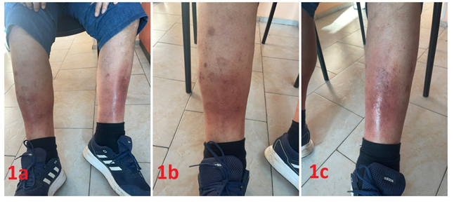
Figures 1a-1c: : Bilateral ill-defined erythema and edema on both legs.
Upon admission to our ward, a pulmonologist was consulted, who confirmed that there was still evidence of changes in the lung
parenchyma associated with covid-19. However, the established changes undergo spontaneous resorption and no additional treatment
is required.
During the dermatological examination, erythematous plaques with ill-defined borders were observed bilaterally on the lower legs (Figure 1a-c). There was also a slight increase in local temperature, spontaneous pain and tenderness. Subcutaneous nodules were palpable (Figure 1). Laboratory tests have been performed, which show data on the presence of an inflammatory process, as abnormalities were detected in the following values: hemoglobin (HGB) - 100.0 g / l; hematocrit (HCT) - 0.283 l / l; MCV - 73.2 fl; MCH - 25.9 pg; MCHC - 354.0 g / l; RDW-CV - 18.1%; ESR - 62 mm / h; blood - urine - test strip - +++; glucose - 10.56 mmol / l; creatinine - 247.4 μmol / l; urea - 8.9 mmol / l; uric acid - 360.0 μmol / l. During the stay, a punch biopsy was taken for histopathological examination, which showed a lobular and septal panniculitis with predominant histiocytic infiltrate with multinucleated giant cells and lipophages (Figure 2a-2f).Systemic therapy was conducted with ceftriaxone 1g x 2 i.v. and methylprednisolone 40mg i.v. with dose reduction according to a schedule for an initial period of 7 days. Enoxaparine was administered prophylactically at a dose of 0.8ml x 1 s.c. Topical treatment was performed with compresses with potassium permanganate 3 times a day and heparin sodium gel / 1000 IU / g - 2 times a day. The patient was discharged with clinical improvement in the form of a decrease in edema, a slight fading of the erythema and a significant disappearance of the pain. For outpatient treatment, dexamethason 4mg (1 / 2-0-1 / 2) was prescribed for 7 days according to the reduction regimen, esomeprazole 40mg (1-0-1) for 15 days, ciprofloxacin 500mg (1- 0-1) for 5 days , аcetylsalicylic acid 100mg (0-0-1) for 30 days, as well as topically: heparin sodium gel / 1000 IU / g- 2 times a day.
During the dermatological examination, erythematous plaques with ill-defined borders were observed bilaterally on the lower legs (Figure 1a-c). There was also a slight increase in local temperature, spontaneous pain and tenderness. Subcutaneous nodules were palpable (Figure 1). Laboratory tests have been performed, which show data on the presence of an inflammatory process, as abnormalities were detected in the following values: hemoglobin (HGB) - 100.0 g / l; hematocrit (HCT) - 0.283 l / l; MCV - 73.2 fl; MCH - 25.9 pg; MCHC - 354.0 g / l; RDW-CV - 18.1%; ESR - 62 mm / h; blood - urine - test strip - +++; glucose - 10.56 mmol / l; creatinine - 247.4 μmol / l; urea - 8.9 mmol / l; uric acid - 360.0 μmol / l. During the stay, a punch biopsy was taken for histopathological examination, which showed a lobular and septal panniculitis with predominant histiocytic infiltrate with multinucleated giant cells and lipophages (Figure 2a-2f).Systemic therapy was conducted with ceftriaxone 1g x 2 i.v. and methylprednisolone 40mg i.v. with dose reduction according to a schedule for an initial period of 7 days. Enoxaparine was administered prophylactically at a dose of 0.8ml x 1 s.c. Topical treatment was performed with compresses with potassium permanganate 3 times a day and heparin sodium gel / 1000 IU / g - 2 times a day. The patient was discharged with clinical improvement in the form of a decrease in edema, a slight fading of the erythema and a significant disappearance of the pain. For outpatient treatment, dexamethason 4mg (1 / 2-0-1 / 2) was prescribed for 7 days according to the reduction regimen, esomeprazole 40mg (1-0-1) for 15 days, ciprofloxacin 500mg (1- 0-1) for 5 days , аcetylsalicylic acid 100mg (0-0-1) for 30 days, as well as topically: heparin sodium gel / 1000 IU / g- 2 times a day.

Figures 2a-2e: : Lobular and septalpanniculitis showing septal thickening and foci of lobular inflammatory infiltrate.
Figure 2f: Detail of the infiltrate showing histiocytes, multinucleate giant cells and lipophages.
Figure 2f: Detail of the infiltrate showing histiocytes, multinucleate giant cells and lipophages.
We describe a third form of panniculitis (lobular / septal with
a predominance of histiocytes and granulomas) in the world’s
literature of a patient who recently had Covid-19 pneumonia.
The post-covid panniculitis recently described in the literature is characterized mainly by eosinophilic, lymphocytic and partly histiocytic infiltrate, and in terms of infiltrate distribution it is of the lobular type [1]. The pathogenesis of eosinophilic panniculitis is currently unclear and somewhat controversial and includes both the idea of reactive response to a wide range of antigenic stimuli such as: 1) lymphomas, atopic dermatitis, leukocytoclastic vasculitis [4], and disorders as a final result of the disruption of tissue integrity like infections such as 2) HIV, streptococcus, toxocariasis, gnathostoimiasis [5]. Of particular interest are the literature data on drug-induced eosinophilic paniculitis after administration of penicillin, heparin or a serum for hyposensitization in patients with bee allergy [6,7]. The second form of lymphocytic panniculitis, described in the literature after a probable Covid-19 infection, concerns a 5-year-old child and the development of lesions involving the face and the upper limbs [2]. The patient was generally asymptomatic, but had been in contact with those infected with Covid- 19 in the family. His smear for Covid-19 was negative, but the serological test was positive for IgG [2]. The authors of the publication present an interesting hypothesis for upregulation of type 1 IFN activity in patients with Covid-19 infection, for example, which has a key role in the host immune response against viral infections [8]. Of interest is the recently shared hypothesis that panniculitis (in general) could occur in the context of disorders of tissue homeostasis due to the incorporation of inorganic material and subsequent activation of the immune cascade [9]. The patient presented by us is an interesting case of advanced septal / lobular panniculitis, which developed immediately after Covid-19 pneumonia, additionally with suspected bladder cancer who also received enoxaparin subcutaneously once daily for a period of about 10 days. Given the complex pathogenesis of panniculitis in general, it could be discussed in this particular case the presence of a concomitant, possibly coincidental, stasis dermatitis (Figure 1) that may have been decompensated together with chronic cardiac failure in the context of Covid-19 infection, thus potentially concurring to trigger the mixed (lobular and septal) panniculitis. Analysis of the present case suggests that the patient’s septal/lobular panniculitis may have a multifactorial nature and can be the result of multiple concomitant factors, namely drugs (enoxaparin / antibiotics), viral-exogenous (Covid-19 pneumonia), paraneoplastic (bladder carcinoma), as well as a mild cardiac decompensation and stasis dermatitis. However, given the timing in the appearance of lesions, Covid-19 infection could be considered a likely major triggering factor. So, even considering the presence of concomitant factors, we favor the thesis that the present case can be probably regarded as a post Covid-19 septal / lobular panniculitis.
The post-covid panniculitis recently described in the literature is characterized mainly by eosinophilic, lymphocytic and partly histiocytic infiltrate, and in terms of infiltrate distribution it is of the lobular type [1]. The pathogenesis of eosinophilic panniculitis is currently unclear and somewhat controversial and includes both the idea of reactive response to a wide range of antigenic stimuli such as: 1) lymphomas, atopic dermatitis, leukocytoclastic vasculitis [4], and disorders as a final result of the disruption of tissue integrity like infections such as 2) HIV, streptococcus, toxocariasis, gnathostoimiasis [5]. Of particular interest are the literature data on drug-induced eosinophilic paniculitis after administration of penicillin, heparin or a serum for hyposensitization in patients with bee allergy [6,7]. The second form of lymphocytic panniculitis, described in the literature after a probable Covid-19 infection, concerns a 5-year-old child and the development of lesions involving the face and the upper limbs [2]. The patient was generally asymptomatic, but had been in contact with those infected with Covid- 19 in the family. His smear for Covid-19 was negative, but the serological test was positive for IgG [2]. The authors of the publication present an interesting hypothesis for upregulation of type 1 IFN activity in patients with Covid-19 infection, for example, which has a key role in the host immune response against viral infections [8]. Of interest is the recently shared hypothesis that panniculitis (in general) could occur in the context of disorders of tissue homeostasis due to the incorporation of inorganic material and subsequent activation of the immune cascade [9]. The patient presented by us is an interesting case of advanced septal / lobular panniculitis, which developed immediately after Covid-19 pneumonia, additionally with suspected bladder cancer who also received enoxaparin subcutaneously once daily for a period of about 10 days. Given the complex pathogenesis of panniculitis in general, it could be discussed in this particular case the presence of a concomitant, possibly coincidental, stasis dermatitis (Figure 1) that may have been decompensated together with chronic cardiac failure in the context of Covid-19 infection, thus potentially concurring to trigger the mixed (lobular and septal) panniculitis. Analysis of the present case suggests that the patient’s septal/lobular panniculitis may have a multifactorial nature and can be the result of multiple concomitant factors, namely drugs (enoxaparin / antibiotics), viral-exogenous (Covid-19 pneumonia), paraneoplastic (bladder carcinoma), as well as a mild cardiac decompensation and stasis dermatitis. However, given the timing in the appearance of lesions, Covid-19 infection could be considered a likely major triggering factor. So, even considering the presence of concomitant factors, we favor the thesis that the present case can be probably regarded as a post Covid-19 septal / lobular panniculitis.
- Dosil VM, Sáez Vicente A, Cortés MM. EosinophilicPanniculitis Associated With COVID-19. ActasDermosifiliogr. 2020;111(9):804-805.
- Pararajasingam A, Goodwin R.GeneralisedPanniculitis as a Post-COVID-19 Presentation in a Child with Aicardi-Goutières Syndrome. Pediatric Dermatology. 2020;37(SUPPL 1):14.
- Requena L, Yus ES. Panniculitis. Part I. Mostly septalpanniculitis. J Am AcadDermatol. 2001;45(2):163-83.
- Rodríguez R, Almagro M, Piñeyro F, Varela L, Jorge B, Del Pozo J, et al. Eosinophilicpanniculitis and insect bite-like eruption in a patient with chronic lymphocytic leukaemia: a spectrum of the same entity. Dermatol Res Pract. 2010;2010:263827.
- Recuero JK, Binda G, Kiszewski AE. Eosinophilicpanniculitis associated with toxocariasis in a child. An Bras Dermatol. 2019;94(2):250-251.
- Masferrer E, Ezquerra G, Escala E, Pujol RM, Arnau A. Eosinophilicpanniculitis triggered by intramuscular penicillin and occupational setting. Allergy. 2011;66(3):436-437.
- Sen BB, Rifaioğlu EN, Ekiz Ö, Özgür T, Akküçük S, İnan MU, et al. Erythematous indurated plaque lesions on the breast. Indian J DermatolVenereolLeprol. 2013;79(6):849.
- Magro CM, M ulvey JJ, Laurence J, Sanders S, Crowson AN, Grossman M, et al. The differing pathophysiologies that underlie COVID-19-associated perniosis and thrombotic retiformpurpura: a case series. Br J Dermatol. 2021;184(1):141-150.
- Lopovok SG, Kolesnikova AO, Egorova ON, Severinova MV, Musatov ID. Panniculitis as a manifestation of metal-associated autoimmune/inflammatory syndrome induced by adjuvants: a case-based review. Rheumatol Int. 2021.