


Research Article
Open Access
The PDGFC CUB Domain Enhances Survival in PDGFC
Mutant Mice
Brian J. Hayes1, Kimberly J. Riehle1,2, Debra G. Gilbertson3, Wendy R. Curtis3, Edward J. Kelly4,
Piper M. Treuting5 and Jean S. Campbell1*
1Department of Pathology, University of Washington, Seattle, WA 98195, USA
2Department of Surgery, University of Washington, Seattle, WA 98195, USA
3Zymogenetics Inc, a Bristol-Myers Squibb Company, Seattle, WA 98102, USA
4Department of Pharmaceutics, University of Washington, Seattle, WA 98195, USA
5Department of Comparative Medicine, University of Washington, Seattle, WA 98195, USA
2Department of Surgery, University of Washington, Seattle, WA 98195, USA
3Zymogenetics Inc, a Bristol-Myers Squibb Company, Seattle, WA 98102, USA
4Department of Pharmaceutics, University of Washington, Seattle, WA 98195, USA
5Department of Comparative Medicine, University of Washington, Seattle, WA 98195, USA
*Corresponding author: Jean S. Campbell, Executive Director of Research and Development, Oncosec Medical, 434 N. 34th St, Suite 100; Seattle,
WA 98103, USA, Tel: +(206) 221-5422; E-mail:
@
Received: May 01, 2015; Accepted: August 27, 2015; Published: September 14, 2015;
Citation: Hayes BJ, Riehle KJ, Gilbertson DG, Curtis WR, Kelly EJ, et al. (2015) The PDGFC CUB domain enhances survival in PDGFC
mutant mice. SOJ Immunol 3(3): 1-11. DOI: http://dx.doi.org/10.15226/soji/3/3/00133
Abstract Top
Platelet-derived growth factor (PDGF) signaling pathways are
necessary for normal development. Here we report a homozygous
pdgf-c mutant mouse that is viable. In this mouse, alternative
splicing gives rise to a truncated transcript containing the entire
coding region of the complement components C1r/C1s sea urchin
EGF bone morphogenetic protein 1 (CUB) domain of PDGFC but
lacking the majority of the Growth Factor Domain (GFD). A mutant
protein is translated from the truncated sequence in vitro. However,
the viability of our homozygous mutant pdgf-c mice depends on the
presence of both pdgfrα alleles.
Keywords: PDGFC; Cleavage, Growth factor; Knockout
Keywords: PDGFC; Cleavage, Growth factor; Knockout
Introduction
The platelet-derived growth factor (PDGF) signal transduction
pathway has important functions in development. Mice lacking
PDGF receptor (PDGFR) -α or –β are embryonic lethal [1,2],
and deletion of pdgf-a, -b, or –c results in perinatal lethality in
homozygous Knockout (KO) animals, though live births are seen
in pdgf-a and pdgf-c KO mice [3-5]. In vitro studies demonstrate
that PDGF-AA, -BB, and -CC induce pdgfrα dimerization, PDGFBB,
and PDGF-DD induce dimerization of pdgfrβ, and -AB, -BB,
and -CC induce αβ -receptor dimerization [6]. As predicted by in
vitro binding studies, pdgf-b KO mice have a phenotype similar
to pdgf-b KO mice, with defects in kidney and hematologic
development [2,4]. Mice lacking PDGFA have a defect in lung
development [3], and interestingly do not phenocopy deletion
of pdgfrα, which causes abnormalities in skeletal development
and neural crest migration [1,7]. On the other hand, pdgf-c
KO animals, referred to as pdgfctm1nagy, have a defect in palate
formation, resulting from abnormal neural crest cell migration
and proliferation [5]. Ding, et al. [5], further reported that pdgf-a,
pdgf-c double KO mice have a phenotype similar to pdgfrα KO
mice, suggesting that PDGF-C is a major activator of pdgfrα signal
transduction in the context of development. PDGF-AA, PDGF-BB, and PDGF-AB are secreted as active
dimers [8], while PDGF-CC and PDGF-DD are secreted as
inactive homodimers requiring extracellular cleavage of an
N-terminal complement components C1r/C1s sea urchin EGF
bone morphogenic protein 1 (CUB) domain to allow receptor
binding (Figure 1a)[9-12]. All PDGFs share a common growth
factor domain (GFD), which is 110 amino acids long and contains
8 conserved cysteines that facilitate intra- and intermolecular
disulfide bonds [13]. PDGF signal transduction is mediated
through the GFD, and mutations of these cysteines and loss of
their disulfide bonds cause a lack of PDGFR signal transduction
[14,15]. Furthermore, mutation of sequences in loops I, II, or III
of the GFD, the domain that physically interacts with PDGFRs,
decreases the GFD's ability to bind PDGFRs [16-18]. Effective
signal transduction by pdgf-c thus should require the conserved
cysteines and loops I-III of the GFD, as well as cleavage of the
CUB domain. We hypothesized that deletion of the majority of
the GFD would thus eliminate receptor binding and downstream
signal transduction. Contrary to our hypothesis, we describe a
homozygous pdgf-c mutant mouse wherein most of the GFD is
indeed deleted, but the expression of the CUB domain in these
mice is sufficient for viability. Viability, however, depends on
genetic background, as has been described for other pdgfrα
mutations [19,20].
Methods
Mouse models
The pdgf-c locus was isolated from a 129S5/SvEvBrd genomic
BAC library. The targeting construct replaced a 5 kb genomic
region on chromosome 3 with an IRES LacZ MC1 Neo cassette.
The homologous arms consisted of a 3 kb 5' fragment and a 2.1 kb
3' fragment. Lex-1 129S5/SvEvBrd ES cells were transfected with
the targeting construct and injected into C57BL/6 blastocysts to
generate chimeras. Chimeras were bred to albino C57BL/6J-Tyrc-2J
mice (The Jackson Laboratory) to generate F1 progeny. F1 hybrids
were crossed to generate initial Mendelian ratios (Table 1). Mice

Figure 1: Design of targeting constructs to inactivate PDGF-C. a) A linear diagram of the PDGF-C protein showing the CUB (orange) and GFD (green)
above the exons that correspond to these protein domains. b) The targeting strategy for PDGFCtm1Lex mice, which removes exons 4 and 5. c) The targeting
strategy for the PDGFCtm1Nagy strain, which removes exon 2 Ding, et al. [5] Abbreviations : splice acceptor (SA), internal ribosomal entry site (IRES),
complement components C1r/C1s sea urchin EGF bone morphogenic protein 1 (CUB), beta galactosidase neomycin ( β GEO), fusion polyprotein (A)
tail( (pA), Lac Z gene encoding beta galactosidase (Lac Z), the enhancer, promoter, and neomycin phosphotransferase (MC1/ HSVtk and Neomycin).
were further backcrossed to a C57BL/6 background six times
to generate the Mendelian ratio for C57BL/6 (Table 1). These
mice can be obtained from the Mutant Mouse Regional Resource
Centers (http://www.mmrrc.org/index.php) supported by the
NIH. Hemizygous pdgfrα mice, pdgfratm11(egfp)sor, were purchased
from The Jackson Laboratory (stock # 007669). Animals were
housed at the University of Washington, which is an Association for the Assessment and Accreditation of Laboratory Animal Care
approved facility, and all experiments were performed with the
University of Washington Institutional Animal Care and Use
Committee approval.
Necropsy and histology
Homozygote pdgfctm1lex mice (n = 4) and WT C57BL/6
Table 1: Heterozygous PDGFCtm1Lex breeding pairs in two backgrounds produce offspring with normal Mendelian distribution.
Cohort |
No. of animals |
+/+ |
pdgfctm1lex/+ |
pdgfctm1lex / pdgfctm1lex |
Chi sq value |
129Sv/EvBrd |
|||||
observed |
99 |
26 |
50 |
23 |
0.19 |
expected |
25 |
50 |
25 |
||
C57BL/6 |
|||||
observed |
265 |
55 |
147 |
63 |
3.7 |
male |
|||||
observed |
146 |
29 |
83 |
34 |
3.1 |
expected |
37 |
73 |
37 |
||
female |
|||||
observed |
119 |
26 |
64 |
29 |
0.83 |
expected |
|
30 |
59 |
30 |
|
|
|
|
|
|
|
Male and female heterozygous pdgfctm1lex/+ mice were bred and the genotypes of the resulting offspring were determined from DNA extracted
from tail snips by PCR. Two different genetic backgrounds, 129SvBrd and C57BL/6, were analyzed. The expected number of pups with a specific
genotype was calculated based on the total number of pups analyzed. Analysis of normal Mendelian distribution was determined by Chi square
analysis with two degrees of freedom. A Chi square number greater than 5.99 is statistically significant.
littermates (n = 4) aged 5 and 8 weeks were euthanized by
CO2 asphyxiation followed by complete necropsy. Blood was
collected for chemistry and complete blood counts performed
by Phoenix Central Laboratories (Everett, WA). Tissues were
collected for histological analysis with immersion fixation in 10%
phosphate-buffered formalin, 4-6 μm sections were made and
stained with hematoxylin and eosin. Tissues examined included:
lungs, esophagus, trachea, liver, kidneys, heart and great vessels,
adrenal glands, gallbladder, brown and white adipose tissue,
exocrine and endocrine pancreas, spleen, thyroid, submandibular,
parotid and sublingual salivary glands, mesenteric lymph nodes,
mesentery, preputial or clitoral glands, skeletal muscle, bladder,
male accessory sex glands, testes or ovaries, haired skin, large
and small intestine, glandular and non-glandular stomach,
uterus and cross section of the head (eyes, middle and internal
ears, oral and nasal cavities, cerebrum, cerebellum, olfactory
lobes, brain stem, pituitary, tongue and teeth). All tissues were
examined by a board-certified Veterinary Pathologist (PMT) and
given a morphological diagnosis where applicable. Skeletons
were processed by eviscerating, removing the brain and as much
muscle tissue and fat as possible, followed by immersion in 95%
ethanol for at least 72 hours, placed in acetone overnight, and
stained the next night at 37°C with a solution of 70% ethanol, 5%
glacial acetic acid, 11.6 μM Alcian blue, and 14.6 μM Alizarin red.
The following day the skeletons were washed in 95% ethanol
and cleared in 2% KOH for 2 days and transferred to 1% KOH
until fully cleared, with the solution refreshed every 2 to 3 days
until soft tissues were cleared, at which point the skeletons were
transferred to 100% glycerol.
Genotyping
Genomic DNA was extracted from mouse tails by incubation
overnight at 55°C in 20 μg/ml Proteinase K (life technologies) in
lysis buffer (200 mM NaCl, 1% W/V SDS, 10mM Tris-HCl, 1 mM EDTA
pH 8.0). Phenol chloroform extraction was performed, followed by ethanol precipitation and resuspension in Tris-HCl 10mM EDTA
1mM pH 7.4 (TE). Polymerase chain reaction was carried out with
0.5 U GemTaq (MGQuest), supplied 5x buffer, and dNTPs to a final
concentration of 0.2mM with primers 5' CCTGGTCAAGCGCTGTGG
3'(200nM), 5' TCTGGATTCATCGACTGTGG 3'(200nM), 5'
ACGGCTAACATGGAGCACG 3' (100 nM). All primers were
designed using Oligo Calc [21]. Cycling conditions were 95°C for
3 min, and 35 cycles of 95°C for 20 sec, 60°C for 30 sec, and 72°C
for 30 sec with a final extension at 72°C for 10 min.
Southern blotting
The targeting construct was verified using standard methods
as described [22]. Briefly, genomic DNA was extracted as
described above and purified DNA was restriction digested using
EcoR1 HF (New England Biolabs) at 37°C overnight. 10μg of
DNA from pdgfctm1lex homozygous, heterozygous, and wild-Type
(WT) mice was loaded onto a Tris-acetate-EDTA (TAE) gel. Following
electrophoresis, the gel was denatured in 0.4M NaOH,
1.5 M NaCl for 10min followed by neutralization in Tris-HCl
with 1.5M NaCl, pH 7.4. DNA was transferred to Hybond N+ (GE
Healthcare) nylon membrane in 10x SSC and cross-linked in a
Stratalinker 1800 (Stratagene). Exonic DNA including exon 6
and the 3' UTR was used as a probe, amplified by 5' CCTTTTAGGTCCTTCAGTTGAGACC
3' and 5 ATCTATGCAAACAGGTTGGAGAAATCC
3'. The probe was labeled with 50 μCi [α-32P] dCTP using
random decamers (DECAprime II, Ambion) to a specific activity
>108 dpm/μg. The blot was prehybridized with Quickhyb (Stratagene)
at 68°C for 30 min and then incubated with the denatured
probe (106dpm/ml) at 68°C for 2 hr. Labeled membranes were
then washed in 2x SSC with 0.1% SDS and 0.2x SSC with 0.1% SDS
at 60°C for 30 min each. The sizes of the digested genomic DNA
were confirmed by comparison with 32P- dCTP end-labeled λHin
III DNA marker (Fermentas).
RT-PCR analysis
Tissues were collected and flash frozen immediately in liquid
nitrogen. RNA was extracted with Trizol (Invitrogen) as per
the manufacturer's instructions, and cDNA synthesized from
0.5 μgRNA. Primers (F1) 5' CTCACGTGTGCTGCTACGAAGG 3'
and (R3) 5' CCCTGCGATTCTCTGCTGCC 3' were used with the
following conditions; 95°C for 3 min, and 35 cycles of 95°C for 15
sec, 58°C for 15 sec, and 72°C for 30 sec with a final extension at
72°C for 10min.
Cloning
WT and pdgfctm1lex cDNA was amplified with primers 5'
GCCCTCGCCCCAGTCAGC 3' and 5' CTCACGTGTGCTGCTACGAAGG
3' to generate pdgf-c and pdgfctm1lex sequences. WT cDNA was
amplified with primers 5' ATGGTGGTGAATCTAAATCTCCTC
3' and 5' CTCACGTGTGCTGCTACGAAGG 3' to generate the GFD
construct. Additionally, a sequence containing a Kozak sequence
and the pdgf-c signal peptide for secretion was added to the 5'
end of the GFD by Amplification (i.e., 5'GCAGAATTGCCACCATGCTCCTCCTCGGCCT-
CCTCCTGCTGACATCTGCCCTGGCCGGCCAAAGAACGGGGACTCGGGCTGAGTCC3').
The pdgf-c, pdgfctm1lex, and GFD sequences were cloned separately into pEF1/myc-His B plasmids (Life Technologies), but retained the termination codons which prevent translation of the myc-His tag. The plasmids were linearized with Mlu I (New England Biolabs).
The pdgf-c, pdgfctm1lex, and GFD sequences were cloned separately into pEF1/myc-His B plasmids (Life Technologies), but retained the termination codons which prevent translation of the myc-His tag. The plasmids were linearized with Mlu I (New England Biolabs).
Transfection and production of conditioned media
(CM)
Cells [HEK293](ATCC® CRL1573™) were maintained at 37°C
95% humidity with 5% CO2. Transfections were performed using
the calcium phosphate method as described [23], and the cells
were subjected to 500 μg/ml G418 (Life Technologies) for two
weeks to select for cells expressing the following constructs:
empty vector (pEF1empty), pEF1PdgfcGFD, pEF1Pdgfcmut, or
pEF1PdgfcFL to produce untagged proteins, pdgf-cGFD, pdgf-cmut
and pdgf-cFL respectively. Confluent cultures were allowed to
condition media for 24 hours, at which point media were collected
and frozen at -80°C. Baby Hamster Kidney (BHK) cells, which do
not express pdgfrα (BHK 570), and BHK cells transfected with
pdgfrα (BHK Rα) were a kind gift from Dr. Daniel Bowen-Pope
[24].
Immunoblot analysis
SDS-PAGE and immunoblotting were carried out as described
[25]. For PDGF-C detection membranes were incubated with goat
anti-mouse PDGF-C antibody (1:1000, AF1447 R&D Systems)
followed by incubation with an anti-goat Horseradish Peroxidase
(HRP) conjugated secondary antibody and detection performed
with ECL (Pierce). For ERK and p-ERK detection, membranes
were first incubated with anti-phospho p44/42 MAPK (1:1000,
9101 Cell Signaling Technology) followed by an anti-rabbit
conjugated secondary antibody. Primary antibody; secondary
antibody complexes were detected with ECL Plus (GE Healthcare)
and imaged with a Storm 840 phosphor imager (GE Healthcare). Immunoblots were then stripped with 2% SDS, 62.5 mM Tris,
pH 6.8 with 7 μl beta-mercaptoethanol per ml. Blots were then
reprobed with anti-ERK1/2 pAb 7884 followed by secondary
and ECL as above. Densitometry analyzes were performed using
Image J software (NIH) and is presented as the relative value of
the phosphorylated protein normalized to the total levels of the
same protein after re-probing.
Results
Homologous recombination targeting strategy
We examined a pdgf-c mutant mouse, pdgfctm1lex, in which
exons 4 and 5 of the GFD are deleted using the targeting strategy
depicted in Figure 1b. Surprisingly, a viable mouse resulted from
this approach; in contrast to the pdgf-c KO mice, which results
in perinatal lethality (pdgfctm1nagy, Figure 1c). Figure 1 shows the
protein domain structure of PDGF-C and compares the different
targeting approaches used to generate the two mutant strains.
pdgfctm1lex mice were derived from a 129S5/SvEvBrd embryonic
stem cell line (Lex1) targeted by homologous recombination to
replace exons 4 and 5. This strategy should eliminate transcription
of the entire GFD (Figure 1b). pdgfctm1nagy mice were derived from
R1 embryonic stem cells targeted by homologous recombination
to replace exon 2 of PDGFC with an SA-IRES-β-geo-pA cassette
[5], preventing transcription downstream of β -geo, including the
CUB and GFD
Verification of recombination
To confirm that our selection cassette was targeted correctly,
we performed Southern blotting on genomic DNA isolated
from homozygous pdgfctm1lex, heterozygous pdgfctm1lex, and WT
littermates. Digestion of genomic DNA with EcoRI should produce
a 12.4 kb band including exons 4, 5, 6, and the 3' untranslated
region (UTR) from WT DNA, and a 6.5 kb band including exon 6
and the 3' UTR in correctly targeted pdgfctm1lex mice (Figure 2a).
A 32P labeled DNA probe with a complementary sequence to exon
6 and the 3' UTR of pdgf-c was used to detect these 12.4 and 6.5
kb bands by hybridization and bands of the expected size were
detected for homozygous pdgfctm1lex, heterozygous pdgfctm1lex, and
WT mice (Figure 2b).
Pdgfctm1lex transcription and translation
3.4 Given our Southern blotting results, we next verified
production of the expected pdgfctm1lex transcript, which should
result from transcription of exons 1, 2, and 3 of pdgf-c (Figure
1c). Attempts to amplify a transcript containing exon 3 and the
internal ribosomal entry site (IRES) (Figure 3a, primers F1 and
R1) or lacZ (Figure 3a, F1 and R2) did not produce a product
(data not shown), suggesting the targeting construct may have
facilitated skipping the KO cassette. RT-PCR of pdgfctm1lex cDNA
using primers F1 and R3 (Figure 3a) generated a 296 bp band,
consistent with a transcript in which exons 1, 2, 3 and 6 remain
(Figure 3b). Sequencing this amplicon revealed that indeed the
region corresponding to WT exons 4 and 5 was removed, but
exon 6 was spliced in frame to the 3' end of exon 3 to create a

Figure 2: Recombination at the pdgf-c locus in the pdgfctm1lex mouse. a) The anticipated results from EcoR1 digestion of genomic DNA from a
wild type mouse and a pdgfctm1lex mouse. d) Southern blot of genomic DNA digested with EcoRI from wild type, heterozygous, and homozygous
pdgfctm1lex mice.

Figure 3: An alternative mRNA is transcribed from the pdgf-c locus in pdgfctm1lex mice, and protein is expressed from this sequence. a) Diagram of the
splicing of wild type and mutant PDGFC with various PCR primers (F1, F2, R1, R2, R3, and R4) used to detect transcripts. b) Amplicons generated by
RT-PCR (F1 and R3) of RNA from kidneys of wild type, heterozygous, and homozygous pdgfctm1lex mice are shown, producing products of 722 bp for
pdgf-c and 296 bp for pdgfctm1lex. c) Immunoblot of conditioned media from transfected HEK293 cells expressing the coding sequences for an empty
vector (empty), the GFD (pdgf-cGFD), pdgfctm1lex (pdgf-cmut), or full-length pdgf-c (pdgf-cFL). The size of DNA ladder (b) and protein standards (c) are
shown to the left of each image.
truncated transcript (Figure 4). To detect the full-length coding
region of PDGFC, we designed primers to anneal to the 5' and
3' UTRs, and found that the pdgfctm1lex transcript retains the
endogenous 5' and 3' UTRs (Figure 3a, F2 and R4) and associated
regulatory elements (data not shown).
The pdgfctm1lex mutant transcript is expected to have an expression pattern similar to WT pdgf-c, given that the selection cassette did not alter the pdgf-c 5' and 3' regulatory elements. The translated protein product of pdgfcMtm1lex is expected to contain the C-terminal 38 amino acids of the GFD and the entire CUB domain, resulting in a predicted molecular weight of 22.5 kDa. We found that HEK293 cells transfected with plasmids expressing the coding regions for full length Pdgf-c (pdgf-cfl), pdgfctm1lex (pdgf-cmut), or pdgf-c GFD (pdgf-cGFD) secrete proteins of the predicted molecular weights into culture media, as determined by immunoblot analysis (Figure 3c). Thus pdgf-cFL, pdgf-cmut and pdgf-cGFD, are all stable protein products.
The pdgfctm1lex mutant transcript is expected to have an expression pattern similar to WT pdgf-c, given that the selection cassette did not alter the pdgf-c 5' and 3' regulatory elements. The translated protein product of pdgfcMtm1lex is expected to contain the C-terminal 38 amino acids of the GFD and the entire CUB domain, resulting in a predicted molecular weight of 22.5 kDa. We found that HEK293 cells transfected with plasmids expressing the coding regions for full length Pdgf-c (pdgf-cfl), pdgfctm1lex (pdgf-cmut), or pdgf-c GFD (pdgf-cGFD) secrete proteins of the predicted molecular weights into culture media, as determined by immunoblot analysis (Figure 3c). Thus pdgf-cFL, pdgf-cmut and pdgf-cGFD, are all stable protein products.
Signal transduction in response to conditioned
medium
To demonstrate that the CUB domain of pdgf-c could an
activates pdgfrα, we assessed ERK phosphorylation in BHK
cells treated with various Conditioned Media (CM). Media
were conditioned by HEK293 cells transfected with plasmids
expressing pdgf-c CUB, pdgf-c GFD, or an empty vector control.
When CM was added to BHK 570 cells, which have no pdgfrα,
no phosphorylation of ERK was detected (data not shown).
When conditioned medium α was cells added to BHK R α ,
which have been transfected with pdgfrα , phosphorylation of
ERK was detected in a time-dependent manner, with maximal
phosphorylation attained after five minutes of exposure
(Figure 5a). Media conditioned by HEK293 cells transfected
with an empty vector control interestingly did show ERK
phosphorylation in BHK R α cells but not in BHK 570 cells, albeit
to a lesser extent than the PDGF-C constructs, suggesting that

Figure 4: Sequence of cDNA from the 296 bp pdgfctm1lex band shown in 3b compared to the consensus mouse sequence for PDGFC, CCDS17425.1. Exon
junctions are indicated above the consensus sequence.
transfection of HEK293 cells induces the release of a molecule
that can signal through pdgfrα When BHK Rα cells are exposed
to increasing ratio of CM to fresh medium we see an increase in
intensity of ERK phosphorylation just as increasing the amount of
recombinant pdgf-c increases ERK phosphorylation (Figure 5b).
CM from pdgf-c GFD and pdgf-c CUB domain transfected HEK 293
cells maintains some ability to induce ERK phosphorylation even
at low CM ratio to fresh medium.
Anatomic characterization
Originally derived on a 129S5/SvEvBrd background, pdgfctm1lex
mice were backcrossed to a C57BL/6 background. Homozygous
F1 C57BL/6 x 129S5/SvEvBrd pdgfctm1lex progeny had a hunched
appearance, and gross skeletal analyzes demonstrated spina
bifida (Figure 5a and 5b), which was confirmed by histology
(data not shown). All other organs were grossly normal. After six
backcrosses, no overt anatomical differences were noted between
homozygous pdgfctm1lex mice and WT C57BL/6 littermates, i.e. the
spina bifida phenotype was rescued. Comprehensive histological
analyses (see methods) conducted on 5 and 8-week old pdgfctm1lex
homozygous mice were unremarkable or showed incidental
lesions, such as dermatitis or mild multifocal extramedullary
hematopoiesis in the liver, which are known to be associated with the C57BL/6 background [26]. Brains of pdgfctm1lex mice
were examined histologically and did not differ from those of
WT littermates; clinical chemistry and blood counts were also
unremarkable (data not shown).
Genotype ratios
We observed normal Mendelian ratios in the offspring
of heterozygote pdgfctm1lex breeding pairs on two genetic
backgrounds (Table 1). We did not observe premature death
in pdgfctm1lex mice up to 12 months of age and did not see sexdependent
differences in viability on the C57BL/6 background, as
has been reported for pdgfctm1nagy mice [20]. Similar to Fredriksson
L, et al. [6] we observed a statistically significant decrease
in body mass in C57BL/6 pdgfctm1lex mice at 3 and 7 months (a
17 to 24% decrease, data not shown). To determine whether
pdgfrα necessary for normal development in pdgfctm1lex mice, we
intercrossed hemizygous pdgfrα mice with heterozygous
pdgfctm1lex mice. Progeny resulting from this cross did not include
mice that were homozygous pdgfctm1lex; hemizygous pdgfrα (Table
2). Neonatal mice homozygous for pdgfctm1lex and hemizygous for
pdgfrα were born but failed to thrive and 100% died prior to the
age of weaning. This result is surprising, as hemizygous pdgfrα
mice are phenotypically normal [1,27]; we thus conclude that
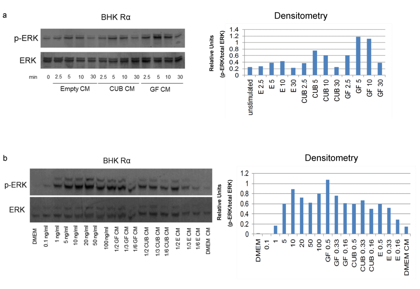
Figure 5: PDGF-C CUB domain stimulates modest ERK phosphorylation in BHK cells expressing PDGFRα. HEK293 cells were transfected with an
empty vector (empty), the CUB domain of PDGF-C (CUB), or the growth factor domain of PDGF-C (GF) and the conditioned media (CM) was transferred
to BHK cell lines expressing PDGFRα.
a) Time-dependent ERK phosphorylation by PDGF-C CUB, PDGF-C GF, or empty vector (E). Immunoblot phospho-ERK1/2 in BHK Rα cells
transfected with pdgfrα after addition of CM. corresponding densitometry measurements are shown to the right.
b) Varying amounts of GF CM, CUB CM, and empty vector CM with non-CM stimulated ERK phosphorylation while DMEM (left lane) and DMEM
conditioned from non-transfected HEK293 cells weakly phosphorylated ERK. Corresponding densitometry measurements are shown to the right.
Note: no phosphorylation of ERK was seen in the parental BHK cells under similar conditions (data not shown).
Table 2: Heterozygous pdgfctm1lex breeding pairs with one copy of pdgfrα do not produce pdgfctm1lex homozygous and hemizygous pdgfrα offspring.
Cohort |
No. of animals |
pdgfc+/+ |
pdgfc+/- |
pdgfc-/- |
pdgfc+/+ |
pdgfc+/- |
pdgfc-/- |
Chi sq value |
observed |
127 |
14 |
27 |
0 |
20 |
50 |
16 |
30 |
expected |
16 |
32 |
16 |
16 |
32 |
16 |
Male and female heterozygous pdgfrtm1lex /+ mice, with a single copy of pdgfrα, were bred, and the genotypes of the resulting offspring were
determined from DNA extracted from tail snips by PCR. The expected number of pups with a specific genotype was calculated based on the total
number of pups analyzed. Analysis of normal Mendelian distribution was determined by Chi squared analysis with five degrees of freedom. A Chi
square number greater than 11.07 is statistically significant. pdgfc-/- represents pdgfctm1lex /tm1lex mice.

Figure 6: Anatomic characterization of pdgfctm1lex mice.
a) 129Sv/EvBrd x C57BL/6 F1 wild type mice have normal thoracic and lumbar vertebrae.
b) Spina bifida in the thoracic and lumbar region (arrow) of pdgfctm1lex homozygous mice.
pdgfctm1lex viability requires two alleles of pdgfrα. Histological
and gross anatomical analysis of these neonatal animals revealed
severe spina bifida encompassing a region from vertebrae T11
to L1 (Figure 6 a-c). Microscopic analysis revealed demyelinated
spinal cords with excessive blood infiltration. Like pdgfctm1nagy
mice, homozygous pdgfctm1lex; hemizygous pdgfrα mice had
palatoschisis. These genetic data indicate that PDGF-Cmut interacts
with pdgfrα in some novel way, as pdgfctm1nagy mice have reduced
viability, while pdgfctm1lex mice have lethality only in conjunction
with hemizygous pdgfrα.
Discussion
PDGF signaling is essential for normal development. In this
study we analyzed a new PDGF-C mutant mouse strain and
demonstrate that the homologous recombination strategy in
this mouse results in an alternatively spliced mutant form of
pdgf-c that lacks over 60% of the RNA encoding the GFD, but
expresses the entire portion of RNA encoding the CUB domain.
This truncated transcript likely results from pre-mRNA splicing,
which is a complicated event relying on proper sequence of both
the 3' and 5' ends of exons, as well as the proper sequence of
the exons themselves [28]. The targeting construct in pdgfctm1lex
used the endogenous 3' splice acceptor sequence of exon 4, but the remainder of exon 4 was replaced with viral and bacterial
sequences (IRES and Lac Z, respectively), which are incompatible
with eukaryotic splicing machinery. The removal of the 3' end of
exon 4 makes splicing at this particular sequence less likely, due
to the inability of U1 to enhance splice site recognition [29]. The
14 bases remaining of the endogenous exon 4 sequence are likely
insufficient to promote splicing, and thus lead to exon skipping in
pdgfctm1lex mice.
The presumed pdgf-c protein that is translated from pdgfctm1lex should not have any GFD function, as paralogs of PDGF-C, PDGF-A and PDGF-B, require cysteines for dimerization and intramolecular and intermolecular folding [14,15]. We hypothesized that the pdgfctm1lex protein product (pdgf-cmut), lacking 6 of the 8 conserved cysteines, would have compromised or absent GFD signal transduction activity. pdgf-cmut is also missing the paralogous loops, I and II of the GFD, which are necessary for receptor binding by the PDGF-C paralog, pdgf-b [16,18]. Loop I has also been shown to be critical for receptor activation by the PDGF paralogs Placental Growth Factor (PlGF) and Vascular Endothelial Growth Factor B (VEGFB), as well as the viral protein VEGFENZ-7 [30,31]. Loop III remains in PDGF-Cmut, but is not expected to bind pdgfrα in the absence of loops I, II and the cysteine required to form loop III. Additionally, PDGFTable
The presumed pdgf-c protein that is translated from pdgfctm1lex should not have any GFD function, as paralogs of PDGF-C, PDGF-A and PDGF-B, require cysteines for dimerization and intramolecular and intermolecular folding [14,15]. We hypothesized that the pdgfctm1lex protein product (pdgf-cmut), lacking 6 of the 8 conserved cysteines, would have compromised or absent GFD signal transduction activity. pdgf-cmut is also missing the paralogous loops, I and II of the GFD, which are necessary for receptor binding by the PDGF-C paralog, pdgf-b [16,18]. Loop I has also been shown to be critical for receptor activation by the PDGF paralogs Placental Growth Factor (PlGF) and Vascular Endothelial Growth Factor B (VEGFB), as well as the viral protein VEGFENZ-7 [30,31]. Loop III remains in PDGF-Cmut, but is not expected to bind pdgfrα in the absence of loops I, II and the cysteine required to form loop III. Additionally, PDGFTable

Figure 7: Developmental divergence between wild-type and homozygous pdgfctm1lex; hemizygous pdgfrα mice.
a) Live P0 pups. Homozygous pdgfctm1lex, hemizygous pdgfrα mice have dark brown linear protrusions at the level of lumbar spine (white arrows).
b) Formalin-fixed homozygous pdgfctm1lex; hemizygous pdgfrα P0 pup with dorsal skin dissected away. Note the protruding and incompletely closed vertebral lamina (arrows) and dark meningomyelocele.
c) And d) Histologic sections through the level of the lumbar spine with kidneys (K) and vertebral body or disc (D) indicated for orientation.
c) Homozygous pdgfctm1lex; hemizygous PDGFRα pup represented in b) the spinal cord and meninges have extensive hemorrhages. The arrow indicates misaligned lamina and there are no dorsal vertebral processes, epaxial skeletal muscles, or abundant subcutaneous tissue covering the spinal cord (contrast to d).
d) Normal thoracic anatomy with proper orientation of vertebral lamina (arrows) and complete enclosure of the spine. Note the skin was removed during dissection.
e) and f) Histological sections of the thoracic spine with lungs (L) indicated for orientation. e) Homozygous pdgfctm1lex; hemizygous PDGFRα pups have severe ascending hemorrhagic myelomalacia.
f) Corresponding cross section of a wild type mouse. Note that the pulmonary hemorrhage and extravasated red blood cells in the meninges are secondary to euthanasia.
g) and h) Cross section of the sinuses and hard palate at the level of the vomeronasal organ (T1).
g) Palatoschisis (cleft palate) is present in the Homozygous pdgfctm1lex; hemizygous PDGFRα mouse (arrowhead).
h) Wild-type mouse.
a) Live P0 pups. Homozygous pdgfctm1lex, hemizygous pdgfrα mice have dark brown linear protrusions at the level of lumbar spine (white arrows).
b) Formalin-fixed homozygous pdgfctm1lex; hemizygous pdgfrα P0 pup with dorsal skin dissected away. Note the protruding and incompletely closed vertebral lamina (arrows) and dark meningomyelocele.
c) And d) Histologic sections through the level of the lumbar spine with kidneys (K) and vertebral body or disc (D) indicated for orientation.
c) Homozygous pdgfctm1lex; hemizygous PDGFRα pup represented in b) the spinal cord and meninges have extensive hemorrhages. The arrow indicates misaligned lamina and there are no dorsal vertebral processes, epaxial skeletal muscles, or abundant subcutaneous tissue covering the spinal cord (contrast to d).
d) Normal thoracic anatomy with proper orientation of vertebral lamina (arrows) and complete enclosure of the spine. Note the skin was removed during dissection.
e) and f) Histological sections of the thoracic spine with lungs (L) indicated for orientation. e) Homozygous pdgfctm1lex; hemizygous PDGFRα pups have severe ascending hemorrhagic myelomalacia.
f) Corresponding cross section of a wild type mouse. Note that the pulmonary hemorrhage and extravasated red blood cells in the meninges are secondary to euthanasia.
g) and h) Cross section of the sinuses and hard palate at the level of the vomeronasal organ (T1).
g) Palatoschisis (cleft palate) is present in the Homozygous pdgfctm1lex; hemizygous PDGFRα mouse (arrowhead).
h) Wild-type mouse.
Cmut lacks arginine 231, which has specifically been shown to be
necessary for cleavage of the CUB domain, and this cleavage has
been suggested to be critical for pdgfrα activation. [9,10]. All of
these structural differences suggest that PDGF-Cmut would not
interact with pdgfrα in the same manner as does PDGF-C. Our
data suggest CUB-mediated activation of pdgfrα and raise the
possibility that the CUB domain interacts with pdgfrα outside of
the ligand binding pocket or perhaps interacts with the receptor
in conjunction with other proteins to influence cell signaling.
CUB domains of proteins other than PDGF-C are involved in a diverse range of functions, including complement activation, developmental patterning, tissue repair, axon guidance and angiogenesis, fertilization, hemostasis, inflammation, neurotransmission, receptor-mediated endocytosis, and tumor suppression [32-34]. CUB domains have even been reported to be involved in cell signaling [35-37]. The human version of the PDGF-C CUB domain stimulates proliferation of human coronary artery smooth muscle cells [38]. Our results show that the PDGF-C CUB domain simulates modest ERK phosphorylation in BHK cells that express pdgfrα, but not in the parental BHK 570 cells, which do not express the receptor. These results suggest that the CUB domain interacts with pdgfrα to transduce intracellular signaling events. This notion is supported by the observations that pups who are homozygous pdgfctm1lex; hemizygous pdgfrα (Table 2) were not viable. The precise mechanisms involved with CUB domain-receptor interactions and subsequent signal transduction events are unknown. Relative to the PDGF-C GFD, however, the CUB domain seems to be less potent in activating the receptor. In vivo, it is possible that in the absence of the GFD, the CUB domain transduces sufficient pdgfrα activity to prevent perinatal lethality in homozygous pdgfctm1lex mice. Genetic background appears to have a profound effect on the PDGF signaling pathway in mice. For example, the Patch mutant mouse (Ph), in which a segment of chromosome 5 including pdgfrα is deleted, has a phenotype that varies with background, as C57BL/6 homozygous Ph mice die earlier in development than do Ph mice on a CBA, or BALB/C background [1].
Likewise, the severity of abnormalities in targeted pdgfrα null mice varies depending on background, with only DBA mice surviving until birth [1,39]. Though our observations may be due in part to strain differences, pdgfctm1lex and pdgfctm1nagy mice appear to have different viabilities and phenotypes. pdgfctm1nagy mice have developmental abnormalities on a 129S1/Sv*129X/ SvJ background, specifically a lethal palate formation defect [5]. These mice are viable on a C57BL/6 background but have brain abnormalities [19,20]. Conversely, pdgfctm1lex mice are viable in two backgrounds, C57BL/6 x 129S5/SvEvBrd and C57BL/6 though two alleles of pdgfrα are necessary for their viability. Mice that are homozygous for pdgfctm1lex and hemizygous for pdgfrα die within days of birth and have severe spina bifida. Our results suggest that the CUB domain of pdgf-c performs a function in pdgfrα signaling. Future experiments are needed to determine whether pdgfctm1nagy mice on C57BL/6 background require two pdgfrα alleles. The availability of pdgfctm1lex mice will facilitate additional genetic screens, either alone or in conjunction with pdgfctm1nagy mice or other mutants, to determine the biological functions of the PDGF-C CUB domain.
CUB domains of proteins other than PDGF-C are involved in a diverse range of functions, including complement activation, developmental patterning, tissue repair, axon guidance and angiogenesis, fertilization, hemostasis, inflammation, neurotransmission, receptor-mediated endocytosis, and tumor suppression [32-34]. CUB domains have even been reported to be involved in cell signaling [35-37]. The human version of the PDGF-C CUB domain stimulates proliferation of human coronary artery smooth muscle cells [38]. Our results show that the PDGF-C CUB domain simulates modest ERK phosphorylation in BHK cells that express pdgfrα, but not in the parental BHK 570 cells, which do not express the receptor. These results suggest that the CUB domain interacts with pdgfrα to transduce intracellular signaling events. This notion is supported by the observations that pups who are homozygous pdgfctm1lex; hemizygous pdgfrα (Table 2) were not viable. The precise mechanisms involved with CUB domain-receptor interactions and subsequent signal transduction events are unknown. Relative to the PDGF-C GFD, however, the CUB domain seems to be less potent in activating the receptor. In vivo, it is possible that in the absence of the GFD, the CUB domain transduces sufficient pdgfrα activity to prevent perinatal lethality in homozygous pdgfctm1lex mice. Genetic background appears to have a profound effect on the PDGF signaling pathway in mice. For example, the Patch mutant mouse (Ph), in which a segment of chromosome 5 including pdgfrα is deleted, has a phenotype that varies with background, as C57BL/6 homozygous Ph mice die earlier in development than do Ph mice on a CBA, or BALB/C background [1].
Likewise, the severity of abnormalities in targeted pdgfrα null mice varies depending on background, with only DBA mice surviving until birth [1,39]. Though our observations may be due in part to strain differences, pdgfctm1lex and pdgfctm1nagy mice appear to have different viabilities and phenotypes. pdgfctm1nagy mice have developmental abnormalities on a 129S1/Sv*129X/ SvJ background, specifically a lethal palate formation defect [5]. These mice are viable on a C57BL/6 background but have brain abnormalities [19,20]. Conversely, pdgfctm1lex mice are viable in two backgrounds, C57BL/6 x 129S5/SvEvBrd and C57BL/6 though two alleles of pdgfrα are necessary for their viability. Mice that are homozygous for pdgfctm1lex and hemizygous for pdgfrα die within days of birth and have severe spina bifida. Our results suggest that the CUB domain of pdgf-c performs a function in pdgfrα signaling. Future experiments are needed to determine whether pdgfctm1nagy mice on C57BL/6 background require two pdgfrα alleles. The availability of pdgfctm1lex mice will facilitate additional genetic screens, either alone or in conjunction with pdgfctm1nagy mice or other mutants, to determine the biological functions of the PDGF-C CUB domain.
Acknowledgment
Grant support: NHLBI, Cardiovascular Pathology training
grant (NIH-HL007312) and HHMI program in Molecular Medicine
(BJH), Herbert Coe Foundation, American Surgical Association
Foundation, and the American College of Surgeons (KJR), Drug
Metabolism Transport and Pharmacogenetics Research Fund in
the School of Pharmacy, University of Washington (EJK), NCI,
mechanisms of PDGF-C induced HCC (NIH-CA127228) (JSC).
Declarations
All animal studies described in this manuscript were
performed with University of Washington Institutional Animal
Care and Use Committee approval. The University of Washington
is an Association for the Assessment and Accreditation of
Laboratory Animal Care approved facility.
Sources of Support
This work was supported by the NHLBI, Cardiovascular
Pathology training grant (NIH-HL007312) and HHMI program
in Molecular Medicine (BJH), Herbert Coe Foundation, American
Surgical Association Foundation, Fred Hutchinson CRC New
Investigator Award and the American College of Surgeons (KJR),
Drug Metabolism Transport and Pharmacogenetics Research
Fund in the School of Pharmacy, University of Washington (EJK),
NCI, Mechanisms of PDGF-C induced HCC (NIH-CA127228) (JSC).
ReferencesTop
- Soriano P. The PDGF alpha receptor is required for neural crest cell development and for normal patterning of the somites. Development. 1997;124(14):2691-2700.
- Soriano P. Abnormal kidney development and hematological disorders in PDGF beta-receptor mutant mice. Genes Dev. 1994;8(16):1888-1896.
- Boström H, Willetts K, Pekny M, et al. PDGF-A signaling is a critical event in lung alveolar myofibroblast development and alveogenesis. Cell. 1996;85(6):863-873.
- Levéen P, Pekny M, Gebre-Medhin S, Swolin B, Larsson E, Betsholtz C, et al. Mice deficient for PDGF B show renal, cardiovascular, and hematological abnormalities. Genes Dev. 1994;8(16):1875-1887.
- Ding H, Wu X, Bostrom H, Kim I, Wong N, Tsoi B, et al. A specific requirement for PDGF-C in palate formation and PDGFR-alpha signaling. Nature genetics. 2004;36(10):1111-1116.
- Fredriksson L, Li H, Eriksson U. The PDGF family: four gene products form five dimeric isoforms. Cytokine & Growth Factor rev. 2004;15(4):197-204.
- Tallquist MD, Soriano P. Cell autonomous requirement for PDGFR alpha in populations of cranial and cardiac neural crest cells. Development 2003;130(3):507-518.
- Betsholtz C, Johnsson A, Heldin CH, Westermark B, Lind P, Urdea MS, et al. cDNA sequence and chromosomal localization of human platelet-derived growth factor A-chain and its expression in tumour cell lines. Nature. 1986;320(6064):695-699.
- Li X, Pontén A, Aase K, Karlsson L, Abramsson A, Uutela M, et al. PDGF-C is a new protease-activated ligand for the PDGF alpha-receptor. Nat Cell Biol. 2000;2(5):302-9.
- Gilbertson DG, Duff ME, West JW, Kelly JD, Sheppard PO, Hofstrand PD, et al. Platelet-derived growth factor C (PDGF-C), a novel growth factor that binds to PDGF alpha and beta receptor. J Biol Chem. 2001;276(29):27406-27414.
- Bergsten E, Uutela M, Li X, Pietras K, Ostman A, Heldin CH, et al. PDGF-D is a specific, protease-activated ligand for the PDGF beta-receptor. Nat Cell Biol. 2001;3(5):512-516.
- LaRochelle WJ, Jeffers M, McDonald WF, Chillakuru RA, Giese NA, Lokker NA, et al. PDGF-D, a new protease-activated growth factor. Nat Cell Biol. 2001;3(5):517-521.
- Reigstad LJ, Sande HM, Fluge Ø, Bruland O, Muga A, Varhaug JE et al. Platelet-derived growth factor (PDGF)-C, a PDGF family member with a vascular endothelial growth factor-like structure. J Biol Chem. 2003;278(19):17114-17120.
- Sauer MK, Donoghue DJ. Identification of nonessential disulfide bonds and altered conformations in the v-sis protein, a homolog of the B chain of platelet-derived growth factor. Mol Cell Biol. 1988;8(3):1011-1018.
- Kenney WC, Haniu M, Herman AC, Arakawa T, Costigan VJ, Lary J, et al. Formation of mitogenically active PDGF-B dimer does not require interchain disulfide bonds. J Biol Chem. 1994;269(16):12351-9.
- Kreysing J, Ostman A, van de Poll M, et al. Identification of three amino acid residues in the B-chain of platelet-derived growth factor with different importance for binding to PDGF alpha- and beta-receptors. FEBS Lett. 1996;385(3):181-184.
- Fenstermaker RA, Poptic E, Bonfield TL, Knauss TC, Corsillo L, Piskurich JF, et al. A cationic region of the platelet-derived growth factor (PDGF) A-chain (Arg159-Lys160-Lys161) is required for receptor binding and mitogenic activity of the PDGF-AA homodimer. J Biol Chem. 1993;268(14):10482-10489.
- Andersson M, Ostman A, Kreysing J, Bäckström G, Van de Poll M, Heldin CH. Involvement of loop 2 of platelet-derived growth factor-AA and -BB in receptor binding. Growth Factors 1995;12(2):159-64.
- Fredriksson L, Nilsson I, Su EJ, Andrae J, Ding H, Betsholtz C, et al. Platelet-derived growth factor C deficiency in C57BL/6 mice leads to abnormal cerebral vascularization, loss of neuroependymal integrity, and ventricular abnormalities. Am J Pathol. 2012;180(3):1136-1144. doi: 10.1016/j.ajpath.2011.12.006.
- Eitner F, Bücher E, van Roeyen C, Kunter U, Rong S, Seikrit C, et al. PDGF-C is a proinflammatory cytokine that mediates renal interstitial fibrosis. J Am Soc Nephrol. 2008;19(2):281-289. doi: 10.1681/ASN.2007030290.
- Kibbe WA. OligoCalc: an online oligonucleotide properties calculator. Nucleic Acids Res. 2007;35(Web Server issue):W43-6.
- Crouthamel MH, Kelly EJ, Ho RJ. Development and characterization of transgenic mouse models for conditional gene knockout in the blood-brain and blood-CSF barriers. Transgenic Res. 2012;21(1):113-130. doi: 10.1007/s11248-011-9512-z.
- Sambrook J, Russell DW. Calcium-phosphate-mediated Transfection of Eukaryotic Cells with Plasmid DNAs. CSH Protoc 2006;2006(1). doi: 10.1101/pdb.prot3871.
- Ferns GA, Sprugel KH, Seifert RA, Bowen-Pope DF, Kelly JD, Murray M, et al. Relative platelet-derived growth factor receptor subunit expression determines cell migration to different dimeric forms of PDGF. Growth Factors. 1990;3(4):315-3.
- Campbell JS, Riehle KJ, Brooling JT, Bauer RL, Mitchell C, Fausto N. Proinflammatory cytokine production in liver regeneration is Myd88-dependent, but independent of Cd14, Tlr2, and Tlr4. J Immunol. 2006;176(4):2522-2528.
- Sundberg JP, Taylor D, Lorch G, Miller J, Silva KA, Sundberg BA et al. Primary follicular dystrophy with scarring dermatitis in C57BL/6 mouse substrains resembles central centrifugal cicatricial alopecia in humans. Vet Pathol. 2011;48(2):513-524. doi: 10.1177/0300985810379431.
- Hamilton TG, Klinghoffer RA, Corrin PD, Soriano P. Evolutionary divergence of platelet-derived growth factor alpha receptor signaling mechanisms. Mol Cell Biol. 2003;23(11):4013-4025.
- Reed R, Maniatis T. A role for exon sequences and splice-site proximity in splice-site selection. Cell. 1986;46(5):681-690.
- Yue BG, Akusjarvi G. A downstream splicing enhancer is essential for in vitro pre-mRNA splicing. FEBS Lett. 1999;451(1):10-14.
- Anisimov A, Leppänen VM, Tvorogov D, et al. The basis for the distinct biological activities of vascular endothelial growth factor receptor-1 ligands. Sci Signal. 2013;6(282):ra52 doi: 10.1126/scisignal.2003905.
- Kiba A, Yabana N, Shibuya M. A set of loop-1 and -3 structures in the novel vascular endothelial growth factor (VEGF) family member, VEGF-ENZ-7, is essential for the activation of VEGFR-2 signaling. J Biol Chem 2003;278(15):13453-13461.
- Thielens NM, Bersch B, Hernandez JF, et al. Structure and functions of the interaction domains of C1r and C1s: keystones of the architecture of the C1 complex. Immunopharmacology. 1999;42(1-3):3-13.
- Nakamura F, Goshima Y. Structural and functional relation of neuropilins. Adv Exp Med Biol. 2002;515:55-69
- Mahoney DJ, Mikecz K, Ali T, et al. TSG-6 regulates bone remodeling through inhibition of osteoblastogenesis and osteoclast activation. J Biol Chem. 2008;283(38):25952-25962. doi: M802138200 [pii] 10.1074/jbc.M802138200.
- Lee HX, Mendes FA, Plouhinec JL, et al. Enzymatic regulation of pattern: BMP4 binds CUB domains of Tolloids and inhibits proteinase activity. Genes Dev. 2009;23(21):2551-2562. doi: 10.1101/gad.1839309.
- Hollway GE, Maule J, Gautier P, et al. Scube2 mediates Hedgehog signalling in the zebrafish embryo. Dev Biol. 2006;294(1):104-118.
- Wu YY, Peck K, Chang YL, et al. SCUBE3 is an endogenous TGF-β receptor ligand and regulates the epithelial-mesenchymal transition in lung cancer. Oncogene. 2011;30(34):3682-3693. doi: onc201185 [pii] 10.1038/onc.2011.85.
- Dijkmans J, Xu J, Masure S, et al. Characterization of platelet-derived growth factor-C (PDGF-C): expression in normal and tumor cells, biological activity and chromosomal localization. The international journal of biochemistry & cell biology. 2002;34(4):414-26.
- McKinnon RD, Waldron S, Kiel ME et al. PDGF alpha-receptor signal strength controls an RTK rheostat that integrates phosphoinositol 3'-kinase and phospholipase Cgamma pathways during oligodendrocyte maturation. J Neurosci. 2005;25(14):3499-3508.

