


Research Article
Open Access
Using a Novel Mucor Indicus CBS 226.29 ET
for Biosynthesis of Gold Nanoparticles and
Applying Them in Nanoremediation of Azo
Dyes
Alshehri ANZ*
Department of Biology, University College in Al-Jummum, Umm Al-Qura University, Makkah, 21955, Saudi Arabia
*Corresponding author: Alshehri ANZ, Department of Biology, University college in Al-Jummum, Umm Al-Qura University, Makkah, 21955, Saudi
Arabia, Tel: +00966563708094; E-mail:
@
Received: 25 January, 2018; Accepted:15 February, 2018; Published: 05 April, 2018
Citation: Alshehri ANZ (2018) Using a Novel Mucor Indicus CBS 226.29 ET for Biosynthesis of Gold Nanoparticles and Applying Them in Nanoremediation of Azo Dyes. SOJ Microbiol Infect Dis 6(2):1-7.
DOI: 10.15226/sojmid/6/2/00192
Abstract Top
Synthesis of nanoparticles by microorganisms may offer an
environmentally friendly and affordable alternative to traditional
physical and chemical methods. The microbial synthesis of
nanoparticles is an approach based on green chemistry which mutually
connects microbial biotechnology and nanotechnology. Fungus of
Mucor Indicus CBS 226.29 ET, in this work was used to synthesize
gold nanoparticles (AuNPs). A surface level Plasmon resonance peak
was observed at 500 nm by UV-bis spectra of AuNPs. Images of AuNP
via transmission electron microscope exhibited various shapes and
dispensability characteristics. The synthesis of AuNPs was optimum
at the conditions 1.5 mM/L of HAuCl4, 0.6 g biomass, and pH range
7-11. The Decolorization of different azo dyes was catalyzed efficiently
with the bio-AuNPs, and a new microbial resource candidate was thus
demonstrated for these AuNPs through green synthesis, along with a
potential bio-AuNP application for Decolorization of azo dyes.
Keywords: Azo dyes; Biosynthesis; Decolorization; Gold nanoparticles; Mucor Indicus
Keywords: Azo dyes; Biosynthesis; Decolorization; Gold nanoparticles; Mucor Indicus
Introduction
Superior characteristics of nanomaterials relative to bulk
materials give them a potential for application in every aspect
of our lives [1]. The high oxidation resistance, stability and
biocompatibility of gold nanoparticles (AuNPs) make them
particularly more attractive than all other nanomaterials [2-4]
many traditional physical and chemical methods are available
for the synthesis of AuNPs, their stability and the concern over
using toxic chemicals during production of AuNPs is considered
problematic [5,6] hence an urgent need exists for developing
an eco-friend process for synthesizing AuNPs. Compared to
chemical and physical methods, biological methods for the
synthesis of AuNPs is more reliable and eco friendly [7]. Several
microorganisms can synthesize AuNPs, including bacteria, fungi
and yeast [7-9]. Fungi offer great advantages for synthesizing
AuNPs, and are therefore considered to be most suitable for
high nanoparticles production [10,11]. In particular, fungi
secrete proteins and secondary metabolites in large quantity
and extracellularly, which can then the separated easily without
recourse to process of downstream [12]. Nevertheless, AuNP
synthesis has only been investigated for fewer than 30 fungal
species [9]. More fungi isolates need to be discovered for the
biosynthesis of AuNPs and to overcome the major challenges
related to shape, size and disparity. These three aforementioned
major challenges have been shown in some reports to be affected
by biomass and substrate concentrations and pH value [13-
15]. The control of the size factor in AuNPs biosynthesis is very
important given that particle size affects the nanomaterials
properties directly. With respect to dyes, the largest chemical
class is azo dyes which exhibit great variety in structure and
color [16], thus serious and huge damage could be occurred to
the environment by disposing effluents containing azo dyes. In
particular, since dyes reflect and absorb sunlight due to their
color content, wastewater can become contaminated which can
thereby disrupt photosynthesis and affect aquatic organisms
growth [17]. Some solutions for treating azo dye pollutants
using green methods have been reported. For instance, some of
microorganisms have been reported to be capable of treating azo
dye pollutants efficiently such as Pseudomonas, Shewanella and
Bacillus [18-20]. The degradation of pollutants could be improved
efficiently by using AuNPs in process of Nanoremediation [21].
Reports on degradation of dyes by biogenic AuNPs are very rare.
In this work, the fungus M. indicus CBS 226.29 ET was used to synthesize AuNPs as eco-friendly method, and an UV-Vis spectrophotometer and Transmission Electron Microscopy (TEM) were used to determine the AuNPs characteristics. The optimum conditions of different parameters for biosynthesis of AuNPs were ascertained, and various azo dyes underwent Decolorization to evaluate catalytic characteristics of the bio- AuNPs being prepared.
In this work, the fungus M. indicus CBS 226.29 ET was used to synthesize AuNPs as eco-friendly method, and an UV-Vis spectrophotometer and Transmission Electron Microscopy (TEM) were used to determine the AuNPs characteristics. The optimum conditions of different parameters for biosynthesis of AuNPs were ascertained, and various azo dyes underwent Decolorization to evaluate catalytic characteristics of the bio- AuNPs being prepared.
Materials and methods
Chemicals and medium
The chemicals used in this study (purity 99%) were
obtained from SIGMA ALDRICH Chemicals, USA. The selection
and cultivation medium was a modified form of Martin Broth
(MMB) at pH 7 consisting of (g/L): 1.0 (NH4)2SO4, 1.0 KH2PO4, 0.5
MgSO4•7H2O, and 1.0 glucose. The solid medium contained 2.0%
(w/v) agar in MMB, and for isolating fungi strain 50 mg/L of each
terramycin and tetracycline were added as selective pressure.
Autoclaving of the media at a temperature of 115°C was done for
15 minutes prior to use.
Isolation and identification of fungus
Soil samples contaminated with heavy metals were collected
from an industrial area in Jeddah-Saudi Arabia the samples were
then diluted serially prior to spreading onto MMB agar plates.
Following a 4 day period of incubation at 30°C, selected colonies
were transferred into 25 mL MMB medium and cultivated for a
further period of 3 days at 30°C and at shaking of 150 r/min. A
line of Lacto Phenol Cotton Blue (LPCB) was then smeared on
a slide and examined under a confocal microscope. A sample of
the fungal strain was picked up using adhesive tape and placed
onto the LPCB for further examination by a confocal microscope
(Leica, Wasteland, Germany). Subsequently, closer examination
of the fungal strain was undertaken utilizing Scanning Electron
Microscopy (SEM, JSM- 5410LV, JEOL Ltd., Japan) as described by
[22]. Following a one month period of screening and enrichment,
a single colony of M. indicus was selected to undergo further
characterization and identification through 26S rRNA gene
sequence analysis. Amplification of the 26S rRNA gene from DNA
of the isolated fungus was achieved by Polymerase Chain Reaction
(PCR), and sequencing of the PCR product was done by Center of
Excellence in Genomic medicine Research-KAU (Saudi Arabia).
The sequence was compared with those on record at GenBank
using the BLAST program, and related sequences were aligned by
using Clustal X (1.8) for constructing a Phylogenetic tree by the
Neighbor -joining method using MEGA (version 5.1) with 1000
bootstrap replicates [23].
Synthesis of AuNPs
Fungus M. indicus CBS 226.29 ET was cultivated over a 2-day
period in 100 mL MMB medium at 30°C and shaking at 150 r/
min. Following incubation, the biomass of M. indicus CBS 226.29
ET was prepared by filtration and washing using sterile distilled
water three times. An appropriate wet biomass concentration
was re-suspended in distilled water prior to further incubation
over a period of 40 hours at 30°C with 1.0 mM/L HAuCl4, and
a control experiment was also carried out without using any
biomass. The following procedure was applied to investigate
influences of concentrations of HAuCl4 and biomass, as well as, pH
values on synthesis of AuNPs: (1) Addition of HAuCl4 in a range
of concentrations (0.1, 0.5, 1.0, 1.5, 2.0, 5.0 mM/L) to the 0.1 g
biomass solutions with a final volume of 4 mL. (2) Suspension of
biomass with a range of mass (0.1, 0.2, 0.3, 0.4, 0.5, 0.6 g) in sterile
distilled water (4 mL) were incubated with 2.0 mM/L of HAuCl4.
(3) Adjustment of the reaction solution to pH (3, 5, 7, 9, 11) was
done by HCl and NaOH. These aforementioned experiments were
carried out for 40 hours at a temperature of 30°C.
Characterization of AuNPs
Examination of AuNPs formation was done by visually
observing color changes of the suspensions before monitoring
with a UV-Vis spectrophotometer (Metash UV-9000, China) to
record spectra ranging between 400 nm and 800 nm at 1 nm
resolution. AuNP morphology was then characterized using a
Transmission Electron Microscope (TEM) (Tecnai G2 Spirit-FEI,
Netherlands) with 120 kV accelerating voltage. Samples of 15 μL
were dropped for the TEM analysis onto a carbon coated copper
grid, which was then dried at room temperature prior to the
analysis.
Decolorization of azo dyes by bio-AuNPs
The M. indicus CBS 226.29 ET and AuNPs were subjected to
ultrasonication process (Ultrasonic Processor CPX 750, USA) to
produce bio-AuNPs for Decolorization of azo dyes. Acid, reactive
and cationic azo dyes were used to demonstrate potential
catalytic activity of the bio-AuNPs in Decolorization of azo dyes.
After addition of dyes to 50 mg/L of the bio-AuNP solution,
and incubation on the shaker at 30°C and 150 r/min, a control
experiment was conducted using ultrasonicated M. indicus CBS
226.29 E cells under identical conditions instead of bio-AuNPs
for Decolorization of 50 mg/L of Acid Brilliant Scarlet GR. The
influence of initial concentration of Acid Brilliant Scarlet GR (25,
50, 100, 200 mg/L) on Decolorization process was examined.
Analytical methods
The Absorption wavelength of azo dyes (λmax) was scanned as
characteristic to monitor the decolorization process using a UV–
Vis spectrophotometer following centrifugation (10,000g) for 5
minutes at varying time intervals. The following equation was
used to calculate the Decolorization proportion (1):
Decolorization (%) = (A0−A1)/=A0 × 100 (1)
Where A0 and A1 represent the dye’s initial and final absorbance respectively
High Performance Liquid Chromatography-Mass Spectrometry (HPLC-MS) was used to detect the intermediate products, which resulted from reduction of Acid Brilliant Scarlet GR by bio-AuNPs, as described earlier (Tan et al., 2013).
Decolorization (%) = (A0−A1)/=A0 × 100 (1)
Where A0 and A1 represent the dye’s initial and final absorbance respectively
High Performance Liquid Chromatography-Mass Spectrometry (HPLC-MS) was used to detect the intermediate products, which resulted from reduction of Acid Brilliant Scarlet GR by bio-AuNPs, as described earlier (Tan et al., 2013).
Results and discussion
Isolation and identification of M. indicus
A fungal strain identified as M. indicus, was isolated from
a sample of contaminated soil with heavy metals taken from
Jeddah-Saudi Arabia. Initial screening of the fungal specimen on
its third day of growth prepared using adhesive tape revealed
highly grown hyphae, sporangium-like structures and spores
under confocal microscope (Figure 1). For finer details, the fungal
strain was examined by SEM. The hyphae, collumella, sporangium,
sporangiospore and collarette (Figure 2) of the strain were
clearly observed. All of these notable features attributed to the
genus Mucor, and based on the morphological specifications, it
was clear that the fungal strain would belong to the genus Mucor.
Morphological specifications are usually sufficient to determine

Figure 1: Images of the isolated fungal strain under confocal microscope show the morphological features.

Figure 2: SEM images of the isolated fungal strain show the morphological features.
a strain’s genus level, but rDNA sequencing is necessary for
distinguishing microorganisms at the level of species. Analysis of
the 26S rRNA gene sequence showed evidence of 100% homology
of the fungus to M. indicus (Genebank accession number CBS
226.29 ET), consequently the identification of the fungus was M. indicus CBS 226.29 ET. The relationship between M. indicus CBS
226.29 ET and other strains was demonstrated in the phylogenic
tree as it is depicted in Figure 3.
Biosynthesis of AuNPs
Biosynthesis of the AuNPs was done by incubating M. indicus
CBS 226.29 ET with HAuCl4. After 4 h of incubation, the reaction
mixture changed from a pale yellowish color to violet (Figure
4). This indicated the formation of gold nanoparticles, and the
color change was indicative of a coherent oscillation of surface
nanoparticles of electron gas resulting in Surface Plasmon
Resonance (SPR) [9]. An absorption peak was observed at 500
nm by UV-vis spectrum of the reaction solution (Figure 5).

Figure 3: Phylogenetic tree of M. indicus CBS 226.29 ET and related species
This was indicative of AuNPs formation [24]. The dispersion of
synthesized AuNPs was characterized by a diversity of shapes,
as shown by the TEM images. The predominant shape was the

Figure 4: The pale yellowish reaction mixture A changed to violet color
B, indicating the formation of gold nano particles.

Figure 5: UV–vis spectra of the dispersed AuNPs synthesized by strain
M. indicus CBS 226.29 ET after 40 h incubation, HAuCl4 solution, and
control strain M. indicus CBS 226.29 ET.
sphere, but triangles and hexagons were also observed, and
with respect to size, spherical particles were found to be smaller
relative to the triangles and polygons (Figure 6). As a genus,
the fungal strain M. indicus is known to be capable of secreting
a range of extracellular enzymes and metabolites in varying
amounts. This advantage makes it a highly suitable candidate for
producing metal nanoparticles on an industrial scale [25,26]. The
results also confirm M. indicus to have exemplary ability in the
biosynthesis of AuNPs, which makes them further suitable for
AuNPs biosynthesis as a potential microbial resource.
Effect of HAuCl4 concentrations
Figure 7 depicts the effects of the HAuCl4 concentrations (0.5,
1.5, and 2.0 mM/L) on the synthesis of AuNPs. A violet or pink
color was observed for those suspensions treated with HAuCl4
for 40 h. Absorption peaks were recorded at 500 nm confirming
AuNPs formation [27], and when the concentration was 1.5 mM/L,
a strong Surface Plasmon Resonance (SPR) band was observed at
500 nm. Thus the optimal concentration of HAuCl4 was 1.5 mM/L
for AuNPs biosynthesis. Increasing the HAuCl4 concentration
to 5.0 mM/L resulted the synthesis of gold particles at large
sizes was achieved by attaching on the tube wall. Large particle
formation was likely to be due to a lack of biomolecules necessary
for capping the synthesized nanoparticles and their efficient
stabilization [21]. The use of Verticillium luteoalbum for AuNPs
synthesis resulted in a similar observation. A concentration of
HAuCl4 below 500 mg/L resulted in a particle size of around
20 nm, whereas a concentration above 500 mg/L resulted in a
distribution of the particle size from 50 nm to several hundred
nanometers. Furthermore, the cells exhibited massive particle
aggregates [13].

Figure 6: The TEM image of AuNPs synthesized by strain M. indicus CBS
226.29 ET

Figure 7: UV-Vis spectra of the dispersed AuNPs synthesized under different
concentrations of HAuCl4 (0.1, 0.5, 1.0, 1.5, 2.0, 5.0 mM) in reaction
solutions.
Effect of biomass concentrations
Figure 8 illustrates effects of the biomass concentrations on the
synthesis of AuNPs. The color of all the reactive solutions changed
to purple indicating synthesis of AuNPs at varying biomass
concentrations. When using 0.1 g of biomass, a broadening of
the Surface Plasmon Resonance (SPR) absorbance of reaction
solution was displayed at the maximum wavelength of 500 nm.
This broadness may be attributed to large formations of AuNPs
(triangle, hexagon and sphere) causing a transverse interaction
of radiance [28,29]. Notably, when the biomass concentration
was increased to 0.6 g, a narrowing of the absorbance band was
evident along with a blue shift. This may be explained as higher
biomass concentrations providing more biomolecules thereby
reducing gold ions and allowing for the synthesis of smaller sized
particles, and hence the blue shift in absorption peak levels [30].

Figure 8: UV-Vis spectra of the dispersed AuNPs synthesized under different
concentrations of strain M. indicus CBS 226.29 ET biomass (0.1,
0.2, 0.3, 0.4, 0.5, 0.6 g) in reaction solutions.
Effect of pH
The influence of pH values on synthesis of AuNPs is illustrated
in Figure 9. An improvement in the average of AuNP synthesis
was spotted when increasing the pH value of solutions. In a
condition of extreme alkalinity (pH 12), the color was observed
to change within 2 h. compared to 4 h. when in a condition of
extreme acidity (pH 3). The SPR band was strongly and sharply
defined when in the range pH 7 to 11, but relatively broader at
pH 3. These sharpness and strength at pH 7 to 11, interpreted
as a better performance at synthesis of AuNPs, may be explained
as being due to a higher stability of capping proteins, which the
M. indicus CBS 226.29 ET secreted them under either alkaline or
neutral conditions, whereas under acidic conditions, protonation
of the carboxylic groups may occur [31,15] also observed this
phenomenon, thereby confirming the ability of the cell free
extract of Trichoderma viride to synthesize AuNPs, which took
place at pH 7 and pH 9, but not pH 5.

Figure 9: UV-Vis spectra of the dispersed AuNPs synthesized under different
conditions of pH (3, 5, 7, 9, 11) in reaction solutions.
Application of bio-AuNPs for azo dye Decolorization
Employing of bio-AuNPs in nanoremediation process as
a decolorative to remove azo dyes was conducted. Figure 10
displays the the Decolorization of various azo dyes due to bio-
AuNPs. This includes four acid azo dyes by rate 81% – 96% in
100–120 min, three reactive azo dyes by rate 45% –70% in
100–180 min, and one cation azo dye by rate 39% in 180 min.
The maximum efficiency in Decolorization was for Acid Brilliant
Scarlet GR at 94.7%. This was therefore selected as a model
dye in further experimental investigation. Figure 11 displays
different concentrations of Acid Brilliant Scarlet GR undergoing
Decolorization processes by the bio-AuNPs. The efficiency of Acid
Brilliant Scarlet GR in Decolorization was found to be greater than
90% over 40 min for a dye concentration of 25 mg/L. When this
concentration was increased to 50 mg/L, the high efficiency over
90% in 100 min was retained, but ultrasonicated M. indicus CBS
226.29 ET cells decolored only 4.8% of Acid Brilliant Scarlet GR
(Figure 12). AuNPs may therefore enhance Decolorization greatly
with the use of high catalytic activities. Analysis of Acid Brilliant
Scarlet GR Decolorization byproducts by HPLC-MS revealed
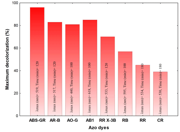
Figure 10: Decolorization of azo dyes (50 mg/L) by bio-AuNPs. Acid
Brilliant Scarlet GR (ABS-GR), Acid Red B (AR-B), Acid Orange G (AO-G),
Acid Black 1 (AB1), Reactive Red X-3B (RR X-3B), Reactive Black (RB),
Reactive Red (RR), Cation Red (CR).

Figure 11: Effects of initial concentration (25, 50, 100, 200 mg/L) of
Acid Brilliant Scarlet GR on Decolorization

Figure 12: The decolorization rate of Acid Brilliant Scarlet GR (50
mg/L) by ultrasonicated M. indicus CBS 226.29 ET cells and bio-AuNPs
(mixture of AuNPs and ultrasonicated M. indicus CBS 226.29 ET cells).
two candidates functioning as Decolorization intermediates,
namely 3, 7-Dihydroxyoctahydronaphthalene- 2,6-dione (I)
corresponding to a mass peak of 215.1658 ([M–H+]), and
naphthol (II) that was emphasized with m/z ion peak of 143.1078
([M–H+]). Previous reports have also detected the same two as
intermediates of Acid Brilliant Scarlet GR degradation [32-34]
indicated that the first step of azo dye degradation was reductive
cleavage of azo groups (–N=N–), however, this study did not
detect 1-aminonaphthylene-2- hydroxy-3, 6-disulfonic acid (III)
as corresponding amines of Acid Brilliant Scarlet GR to which it
was initially transformed. Oxidation of the intermediate (III) was
likely to have been under aerobic conditions to the compound
(I) [33]. The generation of intermediate (II) from product (I)
was likely a result of the removal of two aldehyde groups and
a hydroxyl group. These results indicate a potentially effective
application of bio-AuNPs for azo dye Decolorization.
Conclusions
In this work, an investigation was conducted to synthesize
AuNPs via eco-friendly resource by M. indicus CBS 226.29 ET as a
newly isolated fungal strain. Characteristics of the biosynthesized
AuNPs were ascertained by UV-vis and TEM analysis, and further
experiments were made to investigate the effects of certain
parameters (HAuCl4 concentrations, biomass concentrations
and pH) on AuNPs synthesis to obtain useful data on oriented
biosynthesis of AuNPs. Furthermore, the observations of
nanoremediation of various azo dyes as applications of bio-AuNPs
indicated good potential application of the biogenetic AuNPs in
the Decolorization of azo dyes. This work should provide a more
insight for using fungi as microbial resource in AuNPs synthesis.
ReferencesTop
- Kuang Y, Zhou Y, Chen Z, Megharaj M, Naidu R. Impact of Fe and Ni/Fe nanoparticles on biodegradation of phenol by the strain Bacillus fusiformis (BFN) at various pH values. Bioresour Technol. 2013;136:588-594. doi:10.1016/j.biortech.2013.03.018
- Shukla R, Bansal V, Chaudhary M, Basu A, Bhonde RR, Sastry M. Biocompatibility of gold nanoparticles and their endocytotic fate inside the cellular compartment: a microscopic overview. Langmuir. 2005;21(23):10644-10654. DOI: 10.1021/la0513712
- Bhumkar DR, Joshi HM, Sastry M, Pokharkar VB. Chitosan reduced gold nanoparticles as novel carriers for transmucosal delivery of insulin. Pharm Res. 2007;24(8):1415-1426. DOI: 10.1007/s11095-007-9257-9
- Gong JL, Mullins CB. Surface science investigations of oxidative chemistry on gold. Acc Chem Res. 2009;42(8):1063-1073. DOI: 10.1021/ar8002706
- Narayanan KB, Sakthivel N. Biological synthesis of metal nanoparticles by microbes. Adv Colloid Interf Sci. 2010;156(1-2):1-13. DOI: 10.1016/j.cis.2010.02.001
- Das SK, Das AR, Guha AK. Microbial synthesis of multishaped gold nanostructures. Small. 2010;6(9):1012-1021. doi: 10.1002/smll.200902011
- Zhang X, Yan S, Tyagi RD, Surampalli RY. Synthesis of nanoparticles by microorganisms and their application in enhancing microbiological reaction rates. Chemosphere. 2011; 82(4):489-494. doi:10.1016/j.chemosphere.2010.10.023
- Shedbalkar U, Singh R, Wadhwani S, Gaidhani, S, Chopade BA. Microbial synthesis of gold nanoparticles: current status and future prospects. Adv. Colloid Interf Sci. 2014;209:40-48.
- Kitching M, Ramani M, Marsili E. Fungal biosynthesis of gold nanoparticles: mechanism and scale up. Microb Biotechnol. 2015;8(6):904-917. doi: 10.1111/1751-7915.12151
- Du LW, Xian L, Feng JX. Rapid extra-/intracellular biosynthesis of gold nanoparticles by the fungus Penicillium sp. J Nanopart Res. 2011;13(3):921-930.
- Girard V, Dieryckx C, Job C, Job D. Secretomes: the fungal strike force. Proteomics. 2013;13(3-4):597-608. doi: 10.1002/pmic.201200282
- Mishra A, Kumari M, Pandey S, Chaudhry V, Gupta KC, Nautiyal CS. Biocatalytic and antimicrobial activities of gold nanoparticles synthesized by Trichoderma sp. Bioresour Technol. 2014;166:235-242. doi: 10.1016/j.biortech.2014.04.085
- Gericke, M., Pinches, A. Biological synthesis of metal nanoparticles. Hydrometallurgy. 2006;83(1-4):132-140.
- Pimprikar PS, Joshi SS, Kumar AR, Zinjarde SS, Kulkarni SK. Influence of biomass and gold salt concentration on nanoparticle synthesis by the tropical marine yeast Yarrowia lipolytica NCIM 3589. Colloids Surf. B Biointerfaces. 2009; 74(1):309-316. doi: 10.1016/j.colsurfb.2009.07.040
- Mishra A, Tripathy SK, Yun SI. Bio-synthesis of gold and silver nanoparticles from Candida guilliermondii and their antimicrobial effect against pathogenic bacteria. J Nanosc. Nanotechnol. 2011;11(1):243-248.
- Tony BD, Goyal D, Khanna S. Decolorization of textile azo dyes by aerobic bacterial consortium. Int Biodeterior Biodegrad. 2009;63(4):462-469.
- Champagne PP, Ramsay JA. Dye decolorization and detoxification by laccase immobilized on porous glass beads. Bioresour Technol. 2010;101(7):2230-2235.
- Xu M, Guo J, Kong X, Chen X, Sun G. Fe (III)-enhanced azo reduction by Shewanella decolorationis S12. Appl. Microbiol Biotechnol. 2007;74(6):1342-1349. DOI: 10.1007/s00253-006-0773-z
- Chen XJ, Sun GP, Xu MY. Role of iron in azo reduction by resting cells of Shewanella decolorationis S12. J Appl Microbiol. 2011;110(2):580-586.
- Fang Y, Xu MY, Wu WM, Chen XJ, Sun GP, Guo J, et al. Characterization of the enhancement of zero valent iron on microbial azo reduction. BMC Microbiol. 2015;15(1):85.
- MeenaKumari M, Philip D. Degradation of environment pollutant dyes using phytosynthesized metal nanocatalysts. Spectrochim Acta A. 2015;135:632-638. doi: 10.1016/j.saa.2014.07.037
- Kim KW. Vapor fixation of intractable for simple and versatile scanning electronmicroscope. J Phytopathol. 2008;156:125-128.
- Qu YY, Shi SN, Ma F, Yan B. Decolorization of reactive dark blue KR by the synergism of fungus and bacterium using response surface methodology. Bioresour Technol. 2010;101(21):8016-8023. doi: 10.1016/j.biortech.2010.05.025
- Kalishwaralal K, Deepak V, Pandian SRK, Gurunathan, S. Biological synthesis of gold nanocubes from Bacillus licheniformis. Bioresour Technol. 2009;100(21):5356-5358.
- Vahabi K, Mansoori GA, Karimi S. Biosynthesis of silver nanoparticles by fungus Trichoderma reesei (a route for large-scale production of AgNPs). Insciences J. 2011;(1):65-79. doi:10.5640/insc.010165
- Morin sardin S, Nodet P, Coton E, Jany J. Review: Mucor: a janus-faced fungal genus with human health impact and industrial applications. Fun Bio Reviews. 2017;31:12-32.
- Song JY, Jang HK, Kim BS. Biological synthesis of gold nanoparticles using Magnolia kobus and Diopyros kaki leaf extracts. Process Biochem. 2009;44(10);1133-1138.
- Kreibig U, Genzel L.Optical absorption of small metallic particles. Surf Sci. 1985;156:678-700.
- Sujitha MV, Kannan S. Green synthesis of gold nanoparticles using citrus fruits (Citrus limon, Citrus reticulate and Citrus sinensis) aqueous extract and its characterization. Spectrochim Acta A. 2013;102:15-23.
- Haiss W, Thanh NTK, Aveyard J, Fernig DG. Determination of size and concentration of gold nanoparticles from UV-vis spectra. Anal Chem. 2007;79(11):4215-4221. DOI: 10.1021/ac0702084
- Bastús NG, Comenge J, Puntes V. Kinetically controlled seeded growth synthesis of citrate-stabilized gold nanoparticles of up to 200 nm: size focusing versus Ostwald ripening. Langmuir. 2011;27(17):11098-11105.
- Gomi N, Yoshida S, Matsumoto K, Okudomi M, Konno H, Hisabori T, et al. Degradation of the synthetic dye amaranth by the fungus Bjerkandera adusta Dec 1: inference of the degradation pathway from an analysis of decolorized products. Biodegradation. 2011;22(6):1239-1245. doi: 10.1007/s10532-011-9478-9
- Tan L, Ning S, Zhang X, Shi S. Aerobic decolorization and degradation of azo dyes by growing cells of a newly isolated yeast Candida tropicalis TL-F1. Bioresour Technol. 2013;138:307-313. doi: 10.1016/j.biortech.2013.03.183
- Dos Santos AB, Cervantes FJ, van Lier JB. Review paper on current technologies for decolourisation of textile wastewaters: perspectives for anaerobic biotechnology. Bioresour Technol. 2007;98(12):2369-2385. DOI: 10.1016/j.biortech.2006.11.013

