


Effect of Local Implantation of Composite Biomaterial Loaded with Vascular Endothelial Growth Factor in Healing of Critical-Sized Mandibular Defect- A Pilot Study
Hamid H. Enezei1,2, Azlina A1, Khamis MF1, Rahman RA1, Abdul Razak NH1, Mutum S.S3 and
Samsudin A. R4
1
School of Dental Sciences, Universiti Sains Malaysia Health Campus, 16150 Kelantan, Malaysia
2 College of Dentistry, Anbar University, Ramadi -Iraq, Oral and Maxillofacial Surgery Department.
3 School of Medical Sciences , University Sains Malaysia Health Campus, 16150 Kelantan, Malaysia 4 College of Dentistry, University of Sharjah, Sharjah, United Arab Emirates
2 College of Dentistry, Anbar University, Ramadi -Iraq, Oral and Maxillofacial Surgery Department.
3 School of Medical Sciences , University Sains Malaysia Health Campus, 16150 Kelantan, Malaysia 4 College of Dentistry, University of Sharjah, Sharjah, United Arab Emirates
*Corresponding author: Hamid Hammad Enezei, School of Dental Sciences, Universiti Sains Malaysia Health Campus, 16150 Kelantan, Malaysia, Tel:
+6-0179-295-821; E-mail: drhamed2000@yahoo.com
Received: June 06,2014; Accepted: July 12,2014; Published: July 18,2014
Citation: Enezei HH, Azlina A, Khamis MF, Rahman RA, Abdul Razak NH, et al. (2014) Effect of Local Implantation of Composite
Biomaterial Loaded with Vascular Endothelial Growth Factor in Healing of Critical-Sized Mandibular Defect- A Pilot Study. J Dent
Oral Disord Ther 2(2), 5. DOI: http://dx.doi.org/10.15226/jdodt.2014.00118
Abstract Top
This study was aimed to establish the optimal effective
concentration of VEGF in bone regeneration in vivo. Four male
adult white New Zealand rabbits were equally divided into two
experimental groups, A and B, which were also used as control. One
circular critical size defect 8-mm was created bilaterally in the
mandibular body area of each rabbit. Group A received composite
BCP/FS with 250ng/ml VEGF on one side, and the control side
received BCP/FS only. Group B received composite BCP/FS with
500ng/ml VEGF on one side, and the control side received only
BCP/FS. 4 weeks later, group B showed significant increase in bone
formation compared to that of group A, and least bone formation in
the control.
Keywords: Critical size bone defects; Composite bone graft; Vascular endothelial growth factor
Keywords: Critical size bone defects; Composite bone graft; Vascular endothelial growth factor
IntroductionTop
In human beings, one of the most challenging problems
in oral and maxillofacial surgery is the repair of critical-sized
bone defects (CSBDs) in the mandible that needs attention [1].
Tai K, et al. [1] demonstrated that treatment of CSBDs using
mesenchymal stem cells (MSCs) with suitable biodegradable
scaffolds is feasible. However, this approach is not effective
for CSBDs due to inadequate vascularisation [2]. In order to
encourage the vascularisation process, the cytokines like vascular
endothelial growth factor (VEGF) act as strong promoter agent
of neovascularisation for effective coupling of angiogenesis and
osteogenesis of bone substitutes. The efficacy of VEGF in bone
healing process depends on proper dosage, appropriate delivery
system that has adequate release kinetics and high biological
activity [3]. Thus, this study was aimed to establish the optimal
effective concentration of VEGF in vivo. However, the VEGF has some drawbacks. In high dose administration it has negative
effects on healing process [3]. Furthermore, its half- life is short
when injected into the tissues [4,5]. Considering the drawbacks
and challenge from CSBD, we hypothesized that enriched biphasic
calcium phosphate (BCP) with VEGF and fibrin sealant (FS) can
act as a scaffold with continuous release of low concentration of
VEGF into the area of healing to promote neovascularisation.
Rabbits were sedated before surgery with ketamine xylazine I.M injection, ketamine 35 mg/kg and xylazine 5 mg/kg [6]. Anaesthesia was induced and maintained by administration of 1%-3% sevaflourane mixed with oxygen via a face mask. Rabbits were put in lateral position during surgery, and the heart rate, respiratory rate, pulse oximetry, and temperature of the animals were monitored. Before the operation, the area was shaved and prepared with Betadine, and a subcutaneous injection of 0.5% lidocaine with 1:200,000 epinephrine was given in the line of incision (incision line block). One circular critical-sized trephine defect 8-mm ø was created bilaterally in the mandibular body area (the premolar/ molar region) removing only the lateral bony cortex, trabecular bone, and tooth roots (partial thickness).
Group A received composite BCP/FS loaded with 250ng/ml VEGF, while the other side received only composite without VEGF (control). Group B received composite BCP/FS loaded with 500ng/ ml, while the other side received only composite without VEGF (control). The periosteal flap was repositioned and the wound closed in layers using 4-0 coated Vicryl suture. Postoperatively, each animal was observed closely by a veterinary technician and operator until the regained consciousness at which time, it was transferred to the individual cages. Subcutaneous infusion of 5% glucose water was given with oral orange juice for the initial feeding. Thereafter, the animals were fed normally. In each animal the wound site was debrided daily and cleaned with normal saline and povidon iodine for 7 days. Neomycin local antibiotic cream was applied daily for the first week to the incision site. Intramuscular injection of Baytril (Enrofloxacin), 10mg/kg/day once a day for 7 days followed. Tramadol hydrochloric 2 mg/kg once daily was given for pain relief for 7 days. The weight of the rabbits was recorded daily and any change in the dietary habits or activity was monitored closely. The animals were euthanized at 4 weeks after surgery with an intravenous injection of overdose sodium pentobarbitone (100ml/kg) and the tissues from the defect areas were collected. The samples were fixed in 4% paraformaldehyde for 12 h at room temperature. All samples were scanned for bone formation within the defect site using a Scanco system (µCT 80, Scanco Medical Switzerland). After the µCT scanning, all tissue samples were decalcified in 10% formic acid for tissue processing. All sections were cut and stained with haematoxylin and eosin (H&E) for histomorphometric analysis (Carl Zeiss image analysis system MMI 0684).



Work methodology
All animal procedures were conducted under a protocol
approved by the Universiti Sains Malaysia Animal Ethics
Committee USM (AECUSM) USM/2012/ 1- July- 2012/1- July-
2014), No. of Animal Ethics Approval/USM/ Animal Ethics
Approval/2012/ (81) (419). FS was purchased from Baxter
Healthcare Corporation, Westlake Village, CA- 91362 USA,
US License No. 140. VEGF was purchased from (GIBCO, USA).
Macroporous biphasic calcium phosphate (MBCP) ceramic
granules were synthesized in Ceramic Laboratory, School
of Materials and Mineral Resources, Engineering Campus,
Universiti Sains Malaysia (USM). The preparation of FS and VEGF
was in accordance with the manufacturer's recommendations.
Four male white New Zealand rabbits (Oryctolagus Cuniculus),
aged between 5 and 6 months, and with a mean weight 2.63 kg ±
0.35 kg were divided into two experimental groups A and B. The
experimental rabbits were also used as their own control.Rabbits were sedated before surgery with ketamine xylazine I.M injection, ketamine 35 mg/kg and xylazine 5 mg/kg [6]. Anaesthesia was induced and maintained by administration of 1%-3% sevaflourane mixed with oxygen via a face mask. Rabbits were put in lateral position during surgery, and the heart rate, respiratory rate, pulse oximetry, and temperature of the animals were monitored. Before the operation, the area was shaved and prepared with Betadine, and a subcutaneous injection of 0.5% lidocaine with 1:200,000 epinephrine was given in the line of incision (incision line block). One circular critical-sized trephine defect 8-mm ø was created bilaterally in the mandibular body area (the premolar/ molar region) removing only the lateral bony cortex, trabecular bone, and tooth roots (partial thickness).
Group A received composite BCP/FS loaded with 250ng/ml VEGF, while the other side received only composite without VEGF (control). Group B received composite BCP/FS loaded with 500ng/ ml, while the other side received only composite without VEGF (control). The periosteal flap was repositioned and the wound closed in layers using 4-0 coated Vicryl suture. Postoperatively, each animal was observed closely by a veterinary technician and operator until the regained consciousness at which time, it was transferred to the individual cages. Subcutaneous infusion of 5% glucose water was given with oral orange juice for the initial feeding. Thereafter, the animals were fed normally. In each animal the wound site was debrided daily and cleaned with normal saline and povidon iodine for 7 days. Neomycin local antibiotic cream was applied daily for the first week to the incision site. Intramuscular injection of Baytril (Enrofloxacin), 10mg/kg/day once a day for 7 days followed. Tramadol hydrochloric 2 mg/kg once daily was given for pain relief for 7 days. The weight of the rabbits was recorded daily and any change in the dietary habits or activity was monitored closely. The animals were euthanized at 4 weeks after surgery with an intravenous injection of overdose sodium pentobarbitone (100ml/kg) and the tissues from the defect areas were collected. The samples were fixed in 4% paraformaldehyde for 12 h at room temperature. All samples were scanned for bone formation within the defect site using a Scanco system (µCT 80, Scanco Medical Switzerland). After the µCT scanning, all tissue samples were decalcified in 10% formic acid for tissue processing. All sections were cut and stained with haematoxylin and eosin (H&E) for histomorphometric analysis (Carl Zeiss image analysis system MMI 0684).
Results
The results of the serial postoperative examination revealed
presence of radiological and histological evidence of critical size
bone healing in all treatment groups.
µCT evaluation
The percentage of remaining residual composite was
significantly lower at 4 weeks in BCP/FS loaded with the 500ng/
ml VEGF than in the 250ng/ml of VEGF group as a result of
composite utilization. µCT picture shows radio-lucent area
which is gradually reduced in size and is replaced by opacities
encroaching from the periphery to the centre of the bone defect
with time according to the rate of new bone formation after
surgery (Figure 3). Group B- BCP/FS loaded with 500ng/ml
showed more opacities than group A. The bone regeneration was
increasingly radio-opaque. A clear boundary with the host bone
was observed and different radio-opacities from the periphery
toward the centre of the defect. The amount of newly formed
bone to the total implant area in composite loaded with 500ng/
ml group B was significantly greater than that in the composite
Figure 1: A= BCP granules, B= FS, C= VEGF.
Figure 2: A: CSBD before implantation of composite bone graft. B: CSBD after implantation of composite bone graft
Figure 3: µCT pictures shows a radiolucent zone (halo) surrounding the graft material with minimal size in group B compared to group A and control
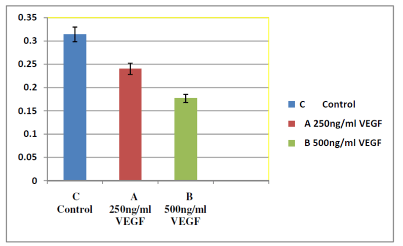
Figure 4: : Mean difference of remaining residual composite (radio-lucent) in group A and B in relation to control group which was measured by µCT less radiolucency mean more new bone formation which appears clear in group B.
Figure 5: Histological presentation at 4 weeks of CSBDs healing in rabbit's mandible in control group, group A and group B (H&E 40x). Group A shows
less new bone formed, with some fibrous tissue, more residual composite (RC) and minimal granulation tissue. Group B shows more scattered new
blood vessels (BV) from defect margin (DM), more granulation tissue (GT), with minimal (RC), marked a new bone formed (NB) in thick layers and
pre-existing bone (PB) appear clearly. In control group a limited amount of new bone formation, more fibrous tissue (FS), inflammatory infiltration
but maximum residual composite (RC) seen.
loaded with 250ng/ml group A and control. Mean difference of
residual composite in group B was less than the mean difference
in group A while mean difference of residual composite was
highest in control group (Figure 4).
Histological and histomorphometrical findings
Histological examination of H&E stained slides demonstrated that more new bone formation occurred in the defect of group
B compared to the defect in group A and control. It was obvious
that composite loaded 500ng/ml VEGF induced more new bone
formation than composite loaded with 250ng/ml four weeks after
implantation. Only little new bone was formed in the composite
loaded with 250ng/ml group. Four weeks after implantation,
most of the composite biomaterial was degraded with more blood

Figure 6: : Histomorphometrical results show the mean difference of groups A and B compared with control. Extensive new bone formation in group
B than in group A and control.
vessels at the periphery and the centre of the defect, the new
bone formed filled most parts of the defect in group B. However,
the defect in group A showed more fibrous tissue than in group
B. It was obvious that composite loaded with 500ng/ml VEGF
induced more new bone formation than composite loaded with
250ng/ml (Figure 5). Histomorphometrical analysis confirm the
histological findings that the amount of newly formed bone to the
total implant area in composite loaded 500ng/ml group B was
significantly greater than that in the composite loaded 250ng/ml
group A and control. For experiment group the mean difference
of group B was higher than the mean difference of group A and
control group meaning that the amount of new bone formation
in group B with composite loaded 500ng/ml VEGF was higher
than the amount of bone formation in group A implanted with
composite loaded with 250ng/ml VEGF. Mean difference of group
A and B was higher than the mean of the control group implanted
with composite only. Percentage of newly formed bone in control
group, composite BCP/FS loaded with 250ng/ml VEGF and
composite BCP/FS loaded with 500ng/ml were 22.5%, 37.5%
and 68.5% respectively as shown in (Figure 6).
Discussion
Existing treatment of CSBDs using autogenous and allograft
procedures are limited and often associated with undesirable
outcomes [7]. Bone xenografts are now considered to be unsuitable
to use as bone graft due to a potential risk of contamination
with viral and bacterial infections, immunogenicity, and finally
host rejection [8]. Composite biomaterials containing calcium
phosphate and polymers (natural or synthetic) have been
developed to address these limitations [9]. FS is a candidate
polymer for use in fabricating composite biomaterials and
delivery vehicle of growth factor for bone regeneration. Numerous
data showed that effects of growth factors on both angiogenesis and bone formation are dose-dependent. The required dose of
growth factor varied with the mode of administration and carrier
type. In general, localized and sustained release of growth factors
allows for lower effective dosage than a single injection [3]. VEGF
has been identified to have positive effects on osteogenesis and
angiogenesis [10,11]. Appropriate dosage and release profiles are
very important for optimized biomolecule delivery. In addition,
the application of excessive dose may provoke adverse effects or
even toxic reactions, such as inhibition of blood vessels and bone
formation [3]. Similarly, slow and sustained release of VEGF can
produce well-functioning blood vessels, whereas uncontrolled
release of VEGF leads to malformed and non functional blood
vessels [12]. Therefore, optimizing and controlling the release of
VEGF in vitro and in vivo is one of the primary concerns in bone
tissue engineering.
Our results are consistent with Hobson's work [13] that VEGF at 500ng/ml generally stimulates a preferable vascularisation. However, very little is known about potential mechanisms that may confine the induction of VEGF within an appropriate magnitude for proper angiogenesis. Because a precise dose of VEGF is critical for vascularisation, disruption of such mechanisms may contribute to pathological angiogenesis [11].
In group B, the regeneration within the defect, characterised by increasing radio-opacity while there is a gradual reduction of the radiolucent zone (halo) surrounding the graft material, indicates more efficient graft resorption when compared to group A and control. The absence of such a radiolucent zone in defect treatment may be indicative of improved new bone formation mediated by the novel bone substitute material. The radiologically detectable sclerotic defect margin present in the present case as well as the decrease in radiolucency can be explained by a summation effect of new bone formation and the bone substitute material still in place. No adverse effect or biological complications has occurred that can be attributed to the biomaterial. Composite in group B showed more abundant blood circulation in the bone defect areas than samples from group A and control because of the physiological function of VEGF as clarified by [14]. This could explain why composite in group B had a higher degradation rate than those of group A and control. In the present study, there was no cartilaginous or fibrocartilaginous tissue in the newly formed bone, and the type of ossification was described as intramembranous origin [15].
Our results are consistent with Hobson's work [13] that VEGF at 500ng/ml generally stimulates a preferable vascularisation. However, very little is known about potential mechanisms that may confine the induction of VEGF within an appropriate magnitude for proper angiogenesis. Because a precise dose of VEGF is critical for vascularisation, disruption of such mechanisms may contribute to pathological angiogenesis [11].
In group B, the regeneration within the defect, characterised by increasing radio-opacity while there is a gradual reduction of the radiolucent zone (halo) surrounding the graft material, indicates more efficient graft resorption when compared to group A and control. The absence of such a radiolucent zone in defect treatment may be indicative of improved new bone formation mediated by the novel bone substitute material. The radiologically detectable sclerotic defect margin present in the present case as well as the decrease in radiolucency can be explained by a summation effect of new bone formation and the bone substitute material still in place. No adverse effect or biological complications has occurred that can be attributed to the biomaterial. Composite in group B showed more abundant blood circulation in the bone defect areas than samples from group A and control because of the physiological function of VEGF as clarified by [14]. This could explain why composite in group B had a higher degradation rate than those of group A and control. In the present study, there was no cartilaginous or fibrocartilaginous tissue in the newly formed bone, and the type of ossification was described as intramembranous origin [15].
Conclusions
The combination of BCP/FS loaded with 500nglml VEGF
significantly accelerates bone healing in CSBDs in vivo. The
results of the present study suggest that BCP and FS are suitable
to act as scaffold in the treatment of CSBD and able to maintain
sustained release of low concentration of VEGF.
Acknowledgement
The authors would like to thank Universiti Sains Malaysia for
the financial support. We also express our gratitude to the staff
of Craniofacial Biology Laboratory and the staff of animal house
(ARASC) for their help
ReferencesTop
- Tai K, Pelled G, Sheyn D, Bershteyn A and Han L. Nanobiomechanics of repair bone regenerated by genetically modified mesenchymal stem cells. Tissue Eng Part A, 2008; 14:1709-1720. doi: 10.1089/ten. tea.2007.0241.
- He X., Dziak R., Yuan X., Mao K., Genco R., Swihart M., et al. BMP2 Genetically Engineered MSCs and EPCs Promote Vascularized Bone Regeneration in Rat Critical-Sized Calvarial Bone Defects. PLoS One, 2013; 8(4):e60473. doi: 10.1371/journal.pone.0060473.
- Bai Y., Yin G., Huang Z., Liao X., Chen X., Yao Y. et al. Localized delivery of growth factors for angiogenesis and bone formation in tissue engineering. Int Immunopharmacol, 2013; 16(2):214-23.
- Lee, K. Y., Peters, M. C. & Mooney. Comparison of vascular endothelial growth factor and basic fibroblast growth factor on angiogenesis in SCID mice. J Controlled Release, 2003; 87(1):49-56.
- Wu C., Zhang Y., Zhou Y., Fan W. & Xiao Y. A comparative study of mesoporous glass/silk and non-mesoporous glass/silk scaffolds: Physiochemistry and in vivo osteogenesis. Acta Biomaterialia, 2012; 7(5):2229-36.
- Daculsi G., Uzel A., Weiss P., Goyenvall E. & Aguado E. Developments in injectable multiphasic biomaterials. The performance of microporous biphasic calcium phosphate granules and hydrogels. J Mater Sci Mater Med, 2010; 21(3):855-61.
- Mehta M., Schmidt-Bleek K., Duda G. N. & Mooney, D. J. Biomaterial delivery of morphogens to mimic the natural healing cascade in bone. Adv Drug Deliv Rev, 2012; 64(12):1257-76. doi: 10.1016/j. addr.2012.05.006.
- Seiichi, Y. The Potential of Tissue Engineering and Regeneration for Craniofacial Bone. Dentistry.
- Osathanon T., Linnes M. L., Rajachar R. M., Ratner B. D., Somerman M. J. & Giachelli C. M. Microporous nanofibrous fibrin-based scaffolds for bone tissue engineering. Biomaterials, 2008; 29(30):4091-99. doi: 10.1016/j.
- Bose, S. & Tarafder, S. Calcium phosphate ceramic systems in growth factor and drug delivery for bone tissue engineering: A review. Acta Biomaterialia, 2012; 8(4):1401-21. doi: 10.1016/j.
- Lu, J. & Tang, M. CTCF-dependent chromatin insulator as a built-in attenuator of angiogenesis. Transcription, 2012; 3(2):73-77. doi: 10.4161/trns
- Kempen D. H., Lu L., Heijink A., Hefferan T. E., Creemers L. B., Maran A., et al. Effect of local sequential VEGF and BMP-2 delivery on ectopic and orthotopic bone regeneration. Biomaterials, 2009; 30(14):2816- 25. doi: 10.1016/j.
- Hobson, M. I. Increased vascularisation enhances axonal regeneration within an acellular nerve conduit. Ann R Coll Surg Engl 2002; 84(1):47.
- Yang, P., Wang, C., Shi, Z., Huang, X., Dang, X., Li, X., et al. rhVEGF 165 delivered in a porous beta-tricalcium phosphate scaffold accelerates bridging of critical-sized defects in rabbit radii. J Biomed Mater Res A, 2010; 92(2):626-40.
- Sencimen M., Aydintug Y., Ortakoglu K., Karslioglu Y., Gunhan O. & Gunaydin Y. Histomorphometrical analysis of new bone obtained by distraction osteogenesis and osteogenesis by periosteal distraction in rabbits. Int. J. Oral Maxillofac. Surg., 2007; 36(3):235-242.

