


2Department of Radiology, East Lancashire Hospital NHS Trust, UK
Methodology: This is a retrospective cohort study of all patients undergoing pancreaticoduodenectomy for pancreatic adenocarcinoma between 2007 and 2012. Patient demographics, pathology data, and resection margin status (R) were analysed. Arterial anatomy based on contrast enhanced computed tomography as per Michels classification was delineated.
Results: Over 5-years, 148 pancreaticoduodenectomies were performed of which 75 were for adenocarcinoma. Fifty-nine patients (78.6%) had a positive resection margin (R1). Descriptive arterial anatomy was available in 72 patients. Fifteen patients (21%) had aberrant anatomy of the hepatic artery. The presence of aberrant arterial anatomy did not have a statistically significant impact on the resection margin status (p=0.67) or involvement of the medial (p=0.34), transection (p=0.22), or posterior (p=0.062) margins. R0 resections demonstrated improved overall survival (p=0.02).
Conclusion: An aberrant hepatic artery is a common finding and should be identified on pre-operative imaging. Aberrant arterial anatomy did not impact on resection margin status. There may be a trend towards posterior tumour margin involvement but this does not affect outcomes in terms of survival.
Key Words: Pancreaticoduodenectomy; Aberrant Hepatic Artery; Resection Margin; Pancreatic Adenocarcinoma; Survival
Michels Classification |
Description |
Frequency (%) |
I |
Normal |
55 |
II |
Replaced LHA from LGA |
10 |
III |
Replaced RHA from SMA |
11 |
IV |
Replaced RHA + LHA |
1 |
V |
Accessory LHA |
8 |
VI |
Accessory RHA |
7 |
VII |
Accessory RHA + LHA |
1 |
VIII |
Replaced RHA + Accessory LHA or Replaced LHA + Accessory RHA |
2 |
IX |
CHA from SMA |
2.5 |
X |
CHA from LGA |
0.5 |
Hiatts Classification |
||
I |
Normal |
75.7 |
II |
Replaced or Accessory LHA |
9.7 |
III |
Replaced or Accessory RHA |
10.6 |
IV |
Replaced or Accessory RHA + Replaced or Accessory LHA |
2.3 |
V |
CHA from SMA |
1.5 |
VI |
CHA from aorta |
0.2 |
The aim of this study was to analyse the frequency and variety of aberrant hepatic arterial anatomy in patients undergoing pancreaticoduodenectomy (PD) for pancreatic adenocarcinoma and whether its presence had an impact on resection margin status and survival.
Statistical analysis was conducted using SPSS Analytic Software (v. 22.0 SPSS Inc., Chicago IL, USA). Continuous variables were compared using Mann-Whitney-U test. Categorical data was compared using Chi-squared or Fishers exact test, as appropriate. Long-term overall survival was taken from the date of surgery to the date of death or the study end date (1st September 2013) and compared using Kaplan-Meier curves. Statistical significance was assessed using the log-rank test. A p-value of < 0.05 was taken as statistically significant.
Aberrant anatomy group (n=15) |
Normal anatomy group |
P-value |
|
Patient Characteristics |
|||
Mean age (SD) |
66.6 (7.2) |
65.2 (9.2) |
0.46 |
Males, n (%) |
8 (53) |
33 (57.8) |
0.77 |
Mean BMI (SD) |
24.8 (4.3) |
25.5 (4) |
0.49 |
Adjuvant Treatment |
|||
Adjuvant Chemotherapy |
10 (80%) |
47 (82%) |
0.94 |
Tumour Characteristics |
|||
>T1, n (%) |
14 (93%) |
56 (98%) |
0.37 |
N1, n (%) |
13 (86%) |
44 (77%) |
0.72 |
Perineural Invasion, n (%) |
12 (80%) |
45 (78%) |
0.98 |
Perivascular Invasion, n (%) |
10 (66%) |
27 (65%) |
1.00 |
Oncological outcomes |
|||
R0 resection, n (%) |
2 (13.3%) |
11 (19.2%) |
0.72 |
Mean Nodal yeild (SD) |
16 (7.3) |
15.4 (6.7) |
0.9 |
Medial/SMV margin involved, n (%) |
9 (60%) |
28 (49%) |
0.21 |
Transection margin involved, n (%) |
4 (26.6%) |
8 (14%) |
0.43 |
Posterior margin involved, n, (%) |
9 (60%) |
17 (30%) |
0.06 |
Michels Classification |
Description |
Frequency, n (% Prevalence) |
I |
Normal |
57 (79%) |
III |
Replaced RHA from SMA |
9 (12.5%) |
V |
Accessory LHA |
2 (2.8%) |
VIII |
Replaced RHA + accLHA or Replaced LHA and accRHA |
2 (2.8%) |
IX |
Replaced CHA from SMA |
1 (1.4%) |
VII |
Accessory RHA and LHA |
1 (1.4%) |
In terms of oncological clearance, 13 patients (18%) had an R0 resection; 11 (19.2%) in the aberrant group and 2 (13.3%) in the normal anatomy group. The relative frequency of R0 resection did not differ significantly between the two groups (p=0.72). Nodal yield during resection also did not differ between the two groups with an average nodal yield of 16 in the aberrant group and 15.4 in the normal group (p=0.90).Of the 59 patients with involved margins, 27 (45%) had more than one resection margin involved. The relative frequency of multiple margin involvement did not differ between the aberrant group (8 of 13, 61.5%) and the normal group (18 of 46, 39%) (p=0.21). The most commonly involved resection margin was the medial SMV resection margin (n=33, 46%) and the posterior resection margin (n=30, 42%). When the individual resection margins were compared between the two groups, there was a trend towards the posterior resection margin being involved more often in the aberrant anatomy group (60% vs. 30%) although this did not reach statistical significance (p=0.062).
Overall median survival was 20 months in the aberrant anatomy group and 17.4 months for the normal anatomy group with no statistically significant difference observed on log-rank test (P=0.84, figure 1). Resection margin status, however, did have an impact on survival where median survival for R0 and R1 resections was 26.8 and 16.8 months respectively (log-rank test, P=0.024, Figure 2).


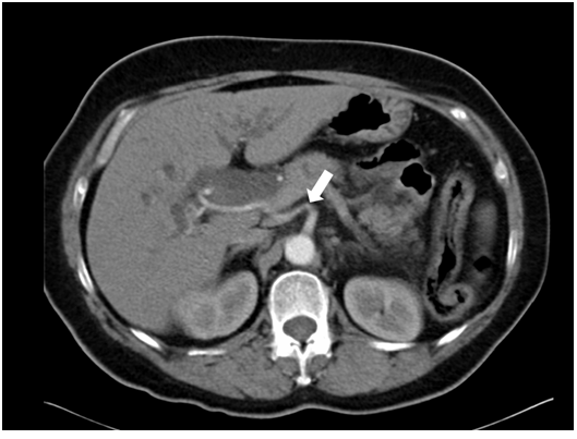
Arising from the SMA, the most common course of anaRHA is along the posterior aspect of the head of the pancreas to the posterolateral border of the hepatoduodenal ligament (Figure 3). Jah et al suggested the exact anatomical course of an aRHA can be divided in three types with the most common (Type 1) taking a posterior route, often within the posterior pancreatic capsule [4]. Less frequently, an aRHA may course through the pancreatic parenchyma (Type 2) or through the SMV groove behind the neck of the pancreas (Type 3). The relatively high frequency of posteriorly coursing vessels could explain why the posterior resection margin was involved more frequently in the aberrant group when compared to the normal group. To our knowledge, this is the first study that has investigated whether an aRHA increases the risk of a specific margin being involved. Although there was a trend towards the posterior resection margin being involved more frequently in the aberrant group, the difference did not attain statistical significance and may be due to the small patient numbers in this subgroup.
The presence of an aRHA can provide several theoretical challenges during surgery. Firstly, intraoperative blood loss due to inadvertent injury, especially when encountered unexpectedly, seems the most obvious. In a series of 143 aRHA, Eshuis et al. described how 5 of the 143 vessels were either accidently ligated or injured. Two of these were repaired primarily, and 1 was reconstructed. Of the 5 patients, two had complications of postoperative haemorrhage and intra abdominal abscess which may have been related to the inadvertent vessel injury [10]. One of the 40 aRHA studied by Kim et al. was injured requiring primary repair and similarly 1 of 43 aRHA discovered by Rammohan et al. was inadvertently ligated but did not result in liver ischaemia [11, 13].
Secondly, once the gastroduodenal artery (GDA) is ligated the arterial supply to the bile duct is heavily reliant on branches of the right hepatic artery which, if sacrificed, renders the proximal common bile duct and therefore bilioenteric anastomosis vulnerable to ischaemia with subsequent breakdown or structuring [14,15]. Finally, attempts to preserve the vessel while ensuring oncological clearance may damage the adventitia increasing the risk of pseudoaneurysm formation and postoperative haemorrhage especially in the setting of a pancreatic fistula or intra-abdominal collections [10,16].
An aberrant hepatic artery can be dealt with safely by sacrifice, avoidance, dissection or transection with vascular reconstruction. Vessel size, tumour proximity and surgeon experience probably all play a role in the decision making process. Whatever the decision, this must follow sound oncological principals while maintaining visceral perfusion. The use of intraoperative Doppler assessment of hepatic flow has been utilised by several authors to aid this decision making process [10, 13]. The vast majority of aRHA can be dissected free and preserved without affecting oncological outcomes, as was the case in our series where 80% of aRHA’s were preserved. In a series of 29 aRHA, Sulpice et al dissected free and preserved 23 vessels, whereas 6 crossed into the tumour and were either sacrificed (n=4) or reconstructed (n=2). Similarly, vessel ligation was deemed necessary for oncological reasons in only 8 of 143 aRHA studied by Eshuis et al. Although considered, reconstruction was not performed. Primary end-to-end or end- to-side anastomosis with the GDA stump or splenic artery are commonly techniques described in the literature [4, 5, 10, 13, 17]. Whereas Stauffer et al. describes using a PTFE jump graft to the GDA in one case, and an interposition gonadal vein graft to the GDA in another [16]. None of the patients in our study required vascular reconstruction.
It is important that aberrant arterial anatomy be identified pre-operatively in order to plan appropriate surgery and minimise the risk of injury. It is also important to consider that CT may not offer adequate sensitivity for diagnosing early tumour involvement and surgeons should modify their surgical approach in the presence of aberrant anatomy, possibly adopting an ‘artery first’ approach. Dissecting the first 3cm of SMA and early retropancreatic right-to-left approach will identify most aberrant right hepatic arteries at which point the vessel can be assessed for tumour involvement and resectability and the need for vascular anastomosis. This was the standard surgical approach in our series. In most instances, the aberrant vessel can be safely preserved but techniques for vascular reconstruction should be adopted if the vessel is of sufficient calibre and must be resected for oncological purposes.
Concordant with other studies, this study has shown that although an aRHA may increase the complexity of pancreaticoduodenectomy, adequate preoperative consideration and intraoperative awareness means its presence should not affect the key objective of clear resection margins and therefore should not impact on survival in patients with pancreatic adenocarcinoma.
- Lin P, Chaikof EL. Embryology, anatomy and surgical exposure of the great abdominal vessels. Surg Clin North Am. 2000;80(1):417-433.
- Michels N. Newer anatomy of the liver and its variant blood supply and collateral circulation. Am J Surg. 1966;112(3):337-347.
- Hiatt J, Gbbay J, Busuttil R. Surgical anatomy of the hepatic arteries in 1000 cases. Ann Surg. 1994;220(1):50-52.
- Jah A, Jamieson N, Huguet E, Praseedom R. The implications of the presence of an aberrant right hepatic artery in patients undergoing a pancreaticoduodenectomy. Surg Today. 2009;39(8):669-674.
- Lee J, Lee Y, Kim C, Moon K, Kim MW. Clinical implications of an aberrant right hepatic artery in patients undergoing pancreaticoduodenectomy. World J Surg. 2009;33(8):1727-1732. doi: 10.1007/s00268-009-0063-x.
- Traverso LW, Freeny PC. Pancreaticoduodenectomy: The importance of preserving hepatic blood flow to prevent biliary fistula. Am Surg. 1989; 55:421-426
- Zhang Y, Frampton AE, Cohen P, Kyriakides CJ, Bong J, Habib NA, et al. Tumor Infiltration in the Medial Resection Margin Predicts Survival After Pancreaticoduodenectomy for Pancreatic Ductal Adenocarcinoma. J Gastrointest Surg. 2012;16(10):1875-1882.
- Verbeke CS, Leitch D, Menon KV, McMahon MJ, Guillou PJ. Redefining the R1 resection in pancreatic cancer. Br J Surg. 2006;93(10):1232-1237.
- Konstantinidis IT, Warshaw AL, Allen JN, Blaszkowsky LS, Castillo CF, Deshpande V, et al. Pancreatic Ductal Adenocarcinoma: Is There a Survival Difference for R1 Resections Versus Locally Advanced Unresectable Tumors? What Is a “True” R0 Resection? Ann Surg. 2013;257(4):731-736. doi: 10.1097/SLA.0b013e318263da2f
- Eshuis WJ, Olde Loohuis KM, Busch ORC, Van Gulik TM, Gouma DJ. Influence of aberrant right hepatic artery on perioperative course and longterm survival after pancreatoduodenectomy. HPB. 2011;13(3):161-167.
- Kim PTW, Temple S, Atenafu EG, Cleary SP, Moulton CA, McGilvray ID, et al. Aberrant right hepatic artery in pancreaticoduodenectomy for adenocarcinoma: impact on resectability and postoperative outcomes. HPB. 2014;16(3):204-211. doi: 10.1111/hpb.12120
- Turrini O, Wiebke EA, Delpero JR, Viret F, Lillemoe KD, van Gulik TM, et al. Preservation of Replaced or Accessory Right Hepatic Artery During Pancreaticoduodenectomy for Adenocarcinoma: Impact on Margin Status and Survival. Journal of Gastrointestinal Surgery. 2010;14(11):1813-1819.
- Rammohan A, Palaniappan R, Pitchaimuthu A, Rajendran K, Senthil Kumar Perumal, Kesavan Balaraman, et al. Implications of the presence of an aberrant right hepatic. World J Gastrointest Surg. 2014;6(1):9-13. doi: 10.4240/wjgs.v6.i1.9
- Schmidt S, Settmacher U, Langrehr J, Neuhaus P. Management and outcomes of patients with combined bile duct and hepatic arterial injuries after laparoscopic cholecystectomy. Surgery. 2004;135(6):613-618.
- Deltenre P, Valla D. Ischemic cholangiopathy. Semin Liver Dis. 2008;28(3):235-246. doi: 10.1055/s-0028-1085092
- Stauffer JA, Bridges MD, Turan N, Nguyen JH, Martin JK, Stauffer J, et al. Aberrant right hepatic arterial anatomy and pancreaticodudenectomy: recognition, prevalence and management. HPB. 2009;11(2):161-165. doi: 10.1111/j.1477-2574.2009.00037.x.
- Shukla PJ, Barreto SG, Kulkarni A, Nagarajan G, Fingerhut A. Vascular anomalies encountered during pancreatoduodenectomy: do they influence outcome? Ann Surg Oncol. 2010;17(1):186-193. doi: 10.1245/s10434-009-0757-1
- Covey AM, Brody LA, Maluccio MA, Getrajdman GI, Brown KT. Variant hepatic arterial anatomy revisited: digital subtraction angiography performed in 600 patients. Radiology. 2002;224(2):542-547.
- Chambers TP, Fishman EK, Bluemke DA, Urban B, Venbrux AC. Identification of the aberrant hepatic artery with axial spiral CT. J Vasc Interv Radiol. 1995;6(6):959-964.

